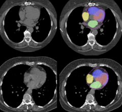
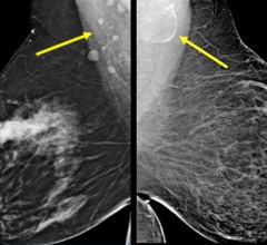
January 11, 2008 - Imaging technologies are helping heart modellers overcome the dilemma of proving that the models really are an accurate representation of the real human heart, as demonstrated in a workshop held recently by the European Science Foundation (ESF).
Computerized in-silico models simulate the real heart and enable possible drugs and therapies to be tested without risk to people.
However, in order to create a realistic model, you need accurate and extensive data from real hearts for calibration.
“Validation of the models is very important, and was raised at the workshop,” said Blanca Rodriguez, scientific coordinator of the ESF workshop, and senior cardiac researcher at Oxford University, Europe’s leading center for cardiac modelling. “One of the problems has been that it is much easier to get experimental data from animals than humans.”
Progress in imaging techniques allows researchers to observe cardiac activity externally without the need for invasive probes. “We are now getting data at very high resolutions and that allows us to model things in more detail, with greatly improved anatomy and structure,” said Rodriguez.
One of the most important diseases being modelled is myocardial ischemia. Studying stem cell therapy to regenerate heart muscle destroyed by disease requires careful testing to eliminate possibly dangerous side effects, such as cancer and disruption of normal heart rhythms, leading to arrhythmia or irregular heart beats. Heart models “could be used to model stem cells’ behavior, and see how they are incorporated into the heart,” said Rodriguez.
Rodriguez recommends that researchers create a standard and robust software infrastructure for sharing heart models and associated data, and to define exactly how to calibrate the models more effectively from experimental data.
For more information: www.esf.org


 July 25, 2024
