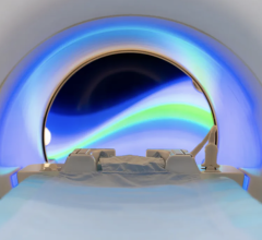
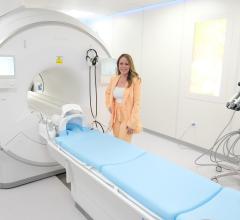
July 23, 2007 - Bringing together the high-quality 3D images of magnetic resonance imaging (MRI) with the intense tumor-killing X-rays of a linear accelerator, scientists at the Alberta Cancer Board are building a prototype that could for the first time enable powerful X-ray beams to become a viable treatment option for liver, stomach and pancreatic cancers, which currently must be treated with surgery, drugs, or internal radioactive seeds in most cases. The hybrid device could also improve results for all cancer patients receiving radiation therapy. Gino Fallone of the Alberta Cancer Board will present this new system at the annual meeting of the American Association of Physicists in Medicine in Minneapolis between July 22-26, 2007.
Radiation therapy is a proven cancer treatment. However, precisely targeting the radiation beam where the tumor is located is still difficult with modern technology. Radiation therapy currently relies on a set of images being taken with radiation (such as X-rays, ultrasound, MRI, PET) and then a complex calculation process to map where the radiation should be targeted.
With this multi-step process, it is difficult to treat local cancers on organs that move, such as the lungs, liver, stomach and pancreas. For other solid tumors, by the time the patient receives radiation their position and sometimes their shape can have changed from the first imaging exams. For this reason, clinicians must allow for a small margin of error. They must apply radiation to a region slightly larger than the tumor to ensure that the entire tumor is treated. Such "treatment margins" damage healthy tissue and can result in more, sometimes serious, side effects. While there are workaround techniques, they can be invasive and problematic.
To overcome the problems of motion and poor tumor definition in X-ray and CT imaging, Alberta Cancer Board researchers Gino Fallone, Marco Carlone and Brad Murray are developing a radiation therapy MRI system using a process called Advanced Real-Time Adaptive RadioTherapy (ART). This process will allow for high-quality, real time MRI imaging at the same time radiation is being administered. MRI provides higher quality images of tumors and organs than X-rays or computed tomography (CT) machines. In addition, it is the only imaging method that can truly provide high-quality, near-real-time 3D images inside the body.
Combining a linear accelerator used for radiation treatments and an MRI is difficult because they function on incompatible scientific and engineering principles. ART overcomes the issue by rotating an MRI machine and a linear accelerator together. The MRI is an "open bore" design, giving patients vastly more room and allowing their heads to be outside the machine for most procedures. The two machines are fixed with respect to each other and rotate in unison around the patient, delivering treatments from all angles. This fixed-system concept reduces electromagnetic interference between the linear accelerator and the MRI and is protected under U.S. and International patents assigned to the Alberta Cancer Board.
The prototype ART is currently under development. A finished prototype is expected by December 2007. When complete, its builders believe will significantly improve the accuracy of radiation treatments for solid tumors, which will result in reduced side effects. More significantly, it will add radiation as a viable treatment option for liver, stomach and pancreatic cancers and could significantly improve treatment for lung and prostate cancers where it is still difficult to administer sufficient radiation doses to obtain a better chance of a cure.
For more information: www.aapm.org


 July 25, 2024


