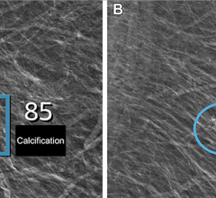
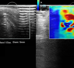
January 30, 2008 – Lesion margins on mammograms and sonograms, calcifications on mammograms, and clinical cancer stage at diagnosis were significantly associated with HER2 status, according to a study conducted to determine if estrogen receptor (ER)-negative human epidermal growth factor receptor type 2 (HER2)-positive and ER-negative HER2-negative breast cancers have distinguishing clinical and imaging features with use of retrospectively identified patients and tissue samples.
In contrast to ER-negative HER2-negative tumors, ER-negative HER2positive tumors were more likely to have spiculated margins (56 percent vs 15 percent), be associated with calcifications (65 percent vs 21 percent), and be detected at a higher cancer stage (74 percent vs 57 percent). Conclusion: Biologic diversity of cancers may manifest in imaging characteristics, and, conversely, studying the range of imaging features of cancers may help refine current molecular phenotypes.
Radiology is a monthly scientific journal devoted to clinical radiology and allied sciences. The journal is edited by Herbert Y. Kressel, M.D., Harvard Medical School, Boston, MA. Radiology is owned and published by the Radiological Society of North America Inc. (RSNA.org/radiologyjnl)
For more information: www.rsna.org


 March 10, 2025


