December 7, 2007 — GE Healthcare has licensed a technique patented by an Eastern Virginia Medical School (EVMS) obstetrician that can automate the acquisition of ultrasound images used by physicians to diagnose fetal heart defects.
GE has licensed the software for exclusive use in its 3D/4D ultrasound systems. Alfred Abuhamad, M.D., chairman of obstetrics and gynecology at EVMS developed the automation protocol, called Sonography based Volume Computer Aided Diagnosis (SonoVCAD).
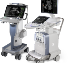
GE has incorporated Abuhamad’s automation protocol in the Voluson E8, the next generation of the GE Voluson ultrasound platform for women’s healthcare. This new 3D/4D ultrasound system includes a number of new tools to help improve clinical workflow, including SonoVCAD.
This paves the way for the future of advanced volume ultrasound and image quality, enabling GE to continue its leadership role in consistently delivering clinically relevant technologies that transform healthcare.
“With some heart defects, infants can die without surgery soon after birth. With an earlier diagnosis months before birth, clinicians and the mother can plan delivery in tertiary care centers with surgeons prepared,” said Abuhamad.
According to the American Heart Association, congenital heart defects rank as the most common birth defect and the number one cause of death during the first year of life.
“Diagnosing defects in the fetal heart requires one of the most challenging diagnostic protocols. It requires a view of the dime-sized heart that shows all four chambers, as well as several precisely angled views of other planes of the heart. If one plane is unobtainable by conventional sonography on the moving fetus, diagnosis is extremely difficult,” Abuhamad said.
Abuhamad’s protocol automates the acquisition of images to display the planes that are needed for a complete ultrasound evaluation of the fetal heart.
“Even for well-trained personnel, manipulation of these planes can be difficult to perform, particularly with relatively complex anatomic organs such as the fetal heart,” said Abuhamad.
This proprietary SonoVCAD technology displays all of the 2D planes, which complies with the recommended standard screening exam of the fetal heart, as outlined by the American Institute of Ultrasound in Medicine (AIUM), the American College of Obstetrics and Gynecology (ACOG), the American College of Radiology (ACR) and the International Society of Ultrasound in Obstetrics and Gynecology (ISUOG). This includes identification of the four-chamber, left outflow tract and right outflow tract views of the fetal heart.
With the software, an ultrasound clinician identifies a standard starting point, for the four-chamber view of the fetal heart. Abuhamad has created algorithms that allow the other planes to be generated from that four-chamber view. Those views allow physicians to identify the type and severity of fetal heart defects.
“SonoVCAD introduces standardization into ultrasound imaging and helps to reduce the risk of operator exam misinterpretation. By simplifying the technical aspects associated with a fetal ultrasound exam, the detection of fetal heart abnormalities should also be enhanced,” said Abuhamad.
For more information: www.gehealthcare.com and www.americanheart.org


 March 19, 2025
