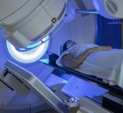
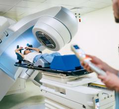
March 7, 2016 — Elekta announced that its Leksell Gamma Knife Icon stereotactic radiosurgery (SRS) system was used for the first time in the United States to treat a metastatic brain tumor. The procedure was conducted on March 1 at Sutter Medical Center, Sacramento Gamma Knife Center in Sacramento, Calif.
The patient, a 52-year-old male from El Dorado Hills, Calif., had previously undergone successful treatment for primary melanoma and for melanoma metastases to his lung. The patient's treatment was planned and guided using a frameless approach. The frameless mask solution is one of several new features of Icon and is integrated with a novel high definition motion management. The system provides accuracy similar to that of frame-based SRS systems while minimizing dose to normal tissue.
"Increasing the precision of frameless cranial SRS is essential for effectively targeting tumor tissue while protecting healthy brain tissue from damage," said Samuel Ciricillo, M.D., medical director of adult and pediatric neurosurgery at Sutter Neuroscience Institute. "The new Gamma Knife system, Icon, now provides the most accurate motion tracking during treatment. Additionally, with Gamma Knife there is a two- to four-fold improvement in sparing normal brain tissue compared to other linear accelerator platforms. These features allow for greater potential to protect patient quality of life both during treatment and after recovery."
Icon is the latest advance in Elekta's Gamma Knife radiosurgery. The system will make cranial SRS available to more patients and improve the efficacy of cranial SRS with fewer side effects. Icon also provides the flexibility for single dose administration or multiple treatment sessions over time, which enables treatment of larger tumor volumes, targets close to critical brain structures, and new or recurring brain metastases.
At the time of SRS, pre-treatment magnetic resonance imaging (MRI) and cone beam computed tomography (CBCT) images are aligned to identify precise coordinates for radiation targeting within the brain. This technology is especially important for patients who undergo multiple treatment sessions. Because the CBCT images are based on fixed structures within the brain, they ensure that dosage and delivery area are calculated correctly for each session, even if the patient's head is in a slightly different position from one treatment session to another.
Ciricillo worked with Sutter Medical Center radiation oncologist Harvey Wolkov, M.D., and physicist Stanley Skubic, Ph.D., on the procedure. They are founding members of the team that started the Sutter Gamma Knife program in 1998.
For more information: www.elekta.com

 August 09, 2024

