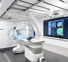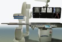
March 18, 2009 - Cardiologists Edward Tadajweski, M.D., and Jeffrey L. Williams, M.D., MS, FACC, are the first in the U.S. to perform 3D imaging of complex cardiac anatomy using GE's Innova 3D imaging system.
On 26 February 2009, they performed 3D reconstructions on two patients' hearts at The Good Samaritan Hospital in Lebanon, PA. The first patient underwent 3D angiography of the coronary sinus to guide a biventricular defibrillator implantation with a left ventricular pacemaker lead. The second patient underwent 3D angiography of the left atrium and pulmonary veins to plan an atrial fibrillation arrhythmia ablation.
“To perform complex interventions and stent placement, deploy [pacemaker or defibrillator] devices or perform atrial fibrillation ablations successfully, you must know exactly where heart anatomy and devices are in relation to one another. Working in a three-dimensional space is critical to better visualize the anatomy before these complex procedures,” said Dr. Tadajweski. “Our state-of-the-art catheterization laboratory allows us to rapidly acquire and reconstruct 3D cardiac anatomy images. It is designed to enhance, but not replace, traditional 2D fluoroscopic imaging.”
The performance of complex cardiac procedures, such as advanced defibrillator placement, structural heart interventions, or atrial fibrillation (arrhythmia) ablation, is facilitated by the visualization of 3D anatomy. Providing 3D views of internal body structures and interventional devices in one image, this state-of-the-art system assists physicians in diagnosis, surgical planning, interventional procedures and treatment follow-up. It permits better management of structural heart disease, streamlines interventional procedures, and minimizes radiation dose to physicians, staff and patients by selecting working views without fluoroscopy.
For more information: www.gshleb.org


 August 14, 2025

