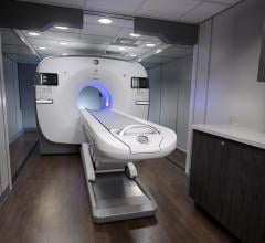
April 19, 2012 — GE Healthcare this week announced it has received U.S. Food and Drug Administration (FDA) clearance of Q.Freeze, one of the positron emission tomography/computed tomography (PET/CT) quantitative imaging technologies designed to enable treatment evaluation earlier in a patient’s cancer treatment.
Q.Freeze combines the quantitative benefits of 4-D phase-matched PET/CT imaging, MotionMatch, into a single static image. By collecting CT and PET data at each phase of the breathing cycle, then matching the data for attenuation correction purposes, Q.Freeze is designed to improve quantitative consistency compared to conventional static PET imaging techniques while facilitating the reading of the 4D PET/CT imaging. None of the acquisition data is wasted, as 100 percent of the counts collected are combined to create a single static image. The goal is an image that has the dual benefit of frozen patient respiratory motion and reduced image noise.
The spark behind the idea — which has been under development since 2006 — was that correcting for lesion movement tied to respiratory motion at the same comfort and dose level as a routine static procedure would support clinicians’ diagnostic confidence. Because patients may not breathe the same way as they might have during their baseline study, Q.Freeze could help enable easy and proper longitudinal response to therapy comparisons.
“GE Healthcare has demonstrated its excellence in addressing one of the biggest clinical challenges in PET/CT: respiratory motion” said Michael Barber, vice president and general manager, molecular imaging, GE Healthcare. “Respiratory motion impacts image clarity and quantification accuracy of lesions in organs subject to respiratory motion, such as the lung, liver and pancreas. Now with Q.Freeze, GE Healthcare is offering an innovative technology that makes patient respiratory motion correction a routine procedure for every scan.”
In a recent study by Cristina Messa, head of the nuclear medicine department, and Luca Guerra, nuclear medicine physician of the Center for Molecular Bioimaging and San Gerado Hospital (HSG-CBM) in Monza, Italy, a patient underwent PET/CT for nodule characterization. Their findings included a comparison of static acquisition, 4-D PET/CT acquisition and Q.Freeze acquisition to determine clear evidence of a metabolic lesion. The Q.Freeze acquisition was able to increase image quality, making the lesion easily identifiable. According to the doctors, this improvement leads to a potential workflow benefit, allowing the physician to review only one set of images free of a patient’s respiratory motion.
Q.Freeze is included in the GE Healthcare Q.Suite, a collection of next-generation capabilities designed to further quantitative PET by generating more consistent standardized uptake value (SUV) readings — enabling clinicians to assess treatment response accurately. During the course of cancer treatment, clinicians traditionally gauge progress by looking for physical change in the size of a tumor, typically using computed tomography (CT) or magnetic resonance (MR). However, with quantitative PET imaging, they are also able to consider a tumor’s metabolic activity. In many cases, metabolic changes in a tumor can be perceived earlier than physical ones; thus quantitative PET can give physicians an earlier view of how well a treatment is working.
For quantitative PET to be effective, consistency of SUV measurements between a patient’s baseline scan and subsequent follow-up scans is critical. Variation can occur throughout the PET workflow, in areas from patient management and biology to equipment protocols and performance. Controlling these variables to increase consistency can improve the clinician’s confidence that an SUV change has true clinical meaning.
“Doctors are seeking quantitative tools, such as Q.Freeze, to obtain reproducible measurements over a longitudinal patient study. Q.Freeze is one of the first element of a suite of tools that may enable doctors to assess confidently biological changes in a patient during a course of treatment, allowing them to quickly and accurately modify treatment regimens,” said Vivek Bhatt, general manager, PET/CT, GE Healthcare. “These tools, ultimately, could potentially contribute to personalized oncology care, increase quality of patient care and reduce wasted expenditure on ineffective treatment.”
For more information: www.gehealthcare.com


 July 30, 2024
