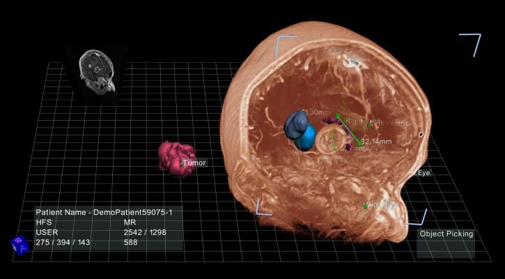
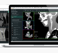
June 2, 2015 — EchoPixel’s True 3D Viewer shows a virtual representation of a patient’s body in open, immersive 3-D. Using stereo glasses, a stylus and a zSpace monitor, doctors can see and manipulate imagery obtained from computed tomography (CT), magnetic resonance (MR) or ultrasound scans as if the objects were on a table in front of them. This helps expand diagnostic and surgical planning.
Capabilities include:
- Measure the diameter of an artery;
- Check for cysts in an organ;
- See exactly where tissue and a tumor intersect;
- Grab, spin and zoom in on the imagery in real time; and
- Identify and separate an object from the rest of the image to get a distinct 3-D view of a tumor, for example.
The high-quality images are powered by the Quadro K4200 graphics card, designed for professional use. The K4200 processes information so fast that doctors can view everything in real time, with no lag in manipulating imagery in 3-D.
True 3D Viewer was recently approved by the U.S. Food and Drug Administration and is in trials with hospitals across the country.
In addition, some companies have an educational version of the viewer. As a patient engagement tool, True 3D Viewer allows doctors to clearly communicate with patients to understand surgeries and improve compliance with treatment plans.
EchoPixel showed True 3D Viewer at the Society for Imaging Informatics in Medicine (SIIM) conference, May 28-30, in Washington, D.C.
For more information: www.echopixeltech.com


 February 01, 2024
