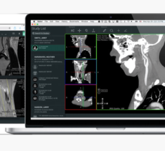
January 8, 2018 — EchoPixel showcased the latest version of True 3D, its interactive, mixed reality software solution at the 103rd Annual Radiological Society of North America (RSNA) meeting, Nov. 26-Dec. 1, 2017, in Chicago. True 3D was featured in the Virtual Reality Showcase in the RSNA Learning Center.
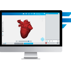
The software offers next-generation surgical planning capabilities. The True 3D Viewer software enables physicians and surgeons to interact with medical images the way they would with physical objects in the real world. The system leverages DICOM image data to create life-size holographic versions of organs, blood vessels and other structures, allowing physicians to rotate, resize, dissect and make incisions to virtual patient-specific anatomy. This capability enables physicians to read images faster, leading to faster diagnoses and provides enhanced pre-operative planning.
Version 1.6.2 expands supported modalities beyond computed tomography (CT) and magnetic resonance imaging (MRI) to include ultrasound with Doppler and C-arm fluoroscopy images. The integration of blood flow, or hemodynamics data, into 3-D representations provides structural and functional data, expanding the clinical utility of ultrasound. For example, this may help better assess implantable device sizing, fibroid and uterine vascularity as well as fetal heart assessment.
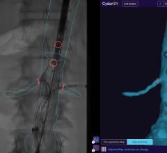
By supporting C-arm views, True 3D software can show a fluoroscopy image in 3-D, as well as use other imaging datasets to create a 3-D digital reconstruction radiograph (DRR), enabling clinicians to better understand the focal point, location and orientation of fluoroscopy images. Visualizing patient anatomy and overlaid fluoroscopy images in the operating room (OR) and cath lab, can help clinicians determine which anatomic structures are in front of or behind the fluoro image plane, as well as better understand where the catheter is in relation to the fluoro plane and patient anatomy.
The latest version of True 3D software also features a new reporting structure specifically designed to help improve the translation of images and expand the surgical planning services radiologists provide surgeons. Key Bookmark Scenes (KBS) files provide a graphical, anatomically meaningful 3-D model that enables the surgeon to visualize complex spatial relationships of the patient's anatomy and pathology.
The virtual patient data is presented in the same view and orientation they would see in the OR. Additionally, the radiologist can create a surgical plan with key step instructions and visualizations. In following the KBS surgical plan, the surgeon can click on the "next step" button and the image data is placed on the current position defined in the plan. This is analogous to using an internet maps app to augment driving directions with 3-D visualizations of key landmarks along the route.
The KBS file can be saved and pushed to the picture archiving and communication system (PACS) for the surgeon to refine the surgical plan and use as a reference during the procedure. The surgeon can interact with the virtual patient's image data, as well as create cross-sections, label key structures and take measurements. For example, the surgeon is able to use interactive tools to visualize different stages for a dissection, surgical procedure or implantable device sizing and implantation.
For more information: www.echopixel.com
Related Content
EchoPixel Announces True 3-D Print Support
EchoPixel True 3D Viewer Gives Surgeons 3-D View Inside the Human Body


 April 18, 2025

