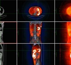
June 19, 2008 - One of the two images selected as the 2008 SNM Image of the Year at SNM 2008 reveals a relapse of neuroendocrine cancer—a malignancy of the interface between the hormonal and nervous systems—including a nodal involvement.
Stefano Fanti, M.D., professor of nuclear medicine at the Policlinico S. Orsola–Università di Bologna in Italy presented the image from a case of a neuroendocrine tumor located in the left middle ear that was referred for restaging.
"The patient was a young male treated by surgery a few months before," said Dr. Fanti. "Conventional imaging, including CT and MR, showed a suspect local relapse. PET/CT confirmed the local relapse, but also demonstrated a nodal involvement, leading to an alteration of his treatment. The finding was subsequently confirmed by CT, and the patient was scheduled for systemic treatment."
The image demonstrates the breadth of molecular imaging and shows how imaging techniques are increasingly being used in combination to provide precise snapshots of both the molecular function and the anatomy of disease in various parts of the human body.
"Using positron emission tomography and computed tomography (PET/CT), one group of physicians was able to see that a suspicious lesion in the left ear was not confined just to that area, but also involved a lymph node. This helped them plan the subsequent treatment," added Dr. Wagner.
For more information: www.snm.org


 July 25, 2024

