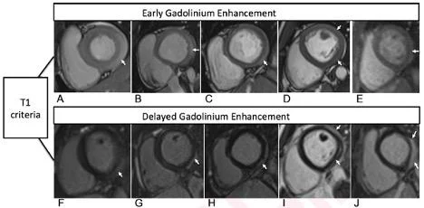
T1-Based Criteria for Myocarditis on Cardiac MRI in Patients With Recent COVID-19 mRNA Vaccination: Early gadolinium enhancement (EGE) compared with precontrast SSFP sequence (not shown) is observed on early postcontrast short-axis SSFP images in (A) 16-year-old male, (B) 17-year-old male, (C) 16-year-old male, and (D) 19-year-old male, and on early postcontrast short-axis perfusion image in (E) 17-year-old male (arrow, A-E). Late gadolinium enhancement (LGE) is also present (arrows) in all 5 patients (F, G, H, I, and J; same patients as in A, B, C, D, and E, respectively). EGE and LGE predominantly affect subepicardium of the inferior, inferolateral, or anterolateral walls. EGE extends beyond confines of LGE in patient shown in (A) and (F) and in (B) and (G). Basilar LV involvement is present in patient shown in (A) and (F), (B) and (G), and (C) and (H). Mid-cavity LV involvement is present in patient shown in (D) and (I) and in (E) and (J).
November 1, 2021 — According to ARRS’ American Journal of Roentgenology (AJR), radiologists need to be cognizant of the association between coronavirus disease (COVID-19) mRNA vaccination and myocarditis, as well as the role of cardiac MRI for assessing suspected myocarditis post-vaccination.
“In this small case series, all patients with myocarditis after COVID-19 vaccination were adolescent males and had a favorable initial clinical course,” explained first author Lydia Chelala from University of Chicago Medicine. Noting that every patient’s cardiac MRI examination showed findings typical of myocarditis from other causes, “late gadolinium enhancement (LGE) persisted in two patients undergoing repeat MRI.”
Chelala and team’s retrospective study included patients who underwent cardiac MRI between May 14, 2021 and June 14, 2021 for suspected myocarditis within 2 weeks of COVID-19 mRNA vaccination—without known prior COVID-19. With clinical presentation, hospital course, and post-discharge events recorded, the cardiac MRI examinations were reviewed in consensus by a cardiothoracic radiologist and cardiothoracic imaging fellow.
Of the 52 patients who underwent cardiac MRI during the study period, Chelala and colleagues identified 5 male patients (age range, 16–19 years; mean age, 17.2 years) who presented within 4 days of the second dose of COVID-19 mRNA vaccine. After mean hospitalization length of 4.8 days, all 5 patients were discharged in stable condition with improved or resolved symptoms. However, two patients underwent repeat cardiac MRI that showed persistent, albeit decreased, LGE.
Acknowledging that their article is the first report to describe additional results of short-term follow-up in this patient population, the authors of this AJR article also conceded, “the observations do not establish causality.”
For more information: www.arrs.org
Related Radiology COVID-19 Content:
Medical AI Models Rely on 'Shortcuts' That Could Lead to Misdiagnosis of COVID-19
CT Provides Best Diagnosis for Novel Coronavirus (COVID-19)
CMS Now Requires COVID-19 Vaccinations for Healthcare Workers by January 4
SNMMI Image of the Year: PET Imaging Measures Cognitive Impairment in COVID-19 Patients
Cardiac MRI Effective in Detecting Asymptomatic, Symptomatic Myocarditis in Athletes
PHOTO GALLERY: How COVID-19 Appears on Medical Imaging
VIDEO: How to Image COVID-19 and Radiological Presentations of the Virus — Interview with Margarita Revzin, M.D.


 July 25, 2024


