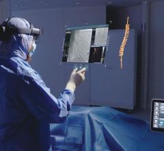
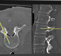
July 29, 2009 - The tubes that power X-ray machines are shrinking, improving the clarity and detail of their Superman-like vision, according to a team of nanomaterial scientists, medical physicists and cancer biologists at the University of North Carolina. The researchers, who presented "Carbon Nanotube Field Emission Based Imaging and Irradiation Technology Development for Basic Cancer Research" on July 28, 2009, at the AAPM 51st Annual Meeting in Anaheim, Calif., have developed new lower-cost X-ray tubes packed with sharp-tipped carbon nanotubes for cancer research and treatment. The tiny technology is being developed to image human breast tissue, laboratory animals and cancer patients under radiotherapy treatment, and to irradiate cells with more control than previously possible with conventional X-ray tubes. The X-ray machine used in a typical hospital today is powered by a "hot" vacuum tube that dates back to the beginning of the 20th century. Inside the tube, a tungsten metal filament - similar to the one that creates light in an incandescent bulb - is heated to a temperature of 1,000 degrees Celsius. The heat releases electrons, which accelerate in the X-ray tube and strike a piece of metal, the anode, creating X-rays. Sha Chang, Otto Zhou, and colleagues that University of North Carolina have developed cold X-ray tubes that replace the tungsten filament with carbon nanotubes packed like blades of tiny grass. Electrons are instantly emitted from the sharp tips of the nanotubes when a voltage is applied. "Think of each nanotube as a lightning rod on top of a building. The high electric field at the tip of the lightning rod draws the electric current from the cloud. Carbon nanotubes emit electrons using a similar principle," said Chang. The group used the nanotubes to build micro-sized scanners and image the interior anatomy of small laboratory animals. Existing X-ray technologies have difficulty compensating for the blur caused by the creature's breathing. Slow mechanical shutters that open and close to block and release the radiation are used to time X-ray pulses to correspond with breath, but their speed is inadequate for small animals because of the creatures' extremely fast breathing and cardiac motion. Chang and Zhou have demonstrated that their carbon nanotubes, which can be turned on and off instantaneously, are fairly easy to synch up to equipment that monitors small animal's breathing or heart rate. The nanotube devices may also improve human cancer imaging and treatment. CT scanners currently in use check for breast cancer by swinging a single large X-ray source around the target to take a thousand pictures over the course of minutes. Using many nanotube X-ray sources lined up in an array instead, breast imaging can be done within few seconds by electronically turning on and off each of the X-ray sources without any physical motion. This fast "tomosynthesis" imaging improves patient comfort and boosts image quality by reducing motion blur. Using 25 simultaneous beams, the team produced images of growths in breast tissue at nearly twice the resolution of commercial scanners on the market. This summer Chang’s team will conduct a clinical test of a first generation nanotube-based imaging system for high-speed image-guided radiotherapy. The research image system is developed by Siemens and Xinray Inc., a joint venture between Siemens and a University of North Carolina startup company Xintech Inc. For more information: www.aapm.org/meetings/09AM/PRAbs.asp?mid=42&aid=11909

 July 18, 2024