Getty Images
August 2, 2023 — Mammography screening supported by artificial intelligence (AI) is a safe alternative to today’s conventional double reading by radiologists and can reduce heavy workloads for doctors. This has now been shown in an interim analysis of a prospective, randomized controlled trial, which addressed the clinical safety of using AI in mammography screening. The trial, led by researchers from Lund University in Sweden, has been published in The Lancet Oncology.
Each year around one million women in Sweden are called to mammography screening. Each screening examination is reviewed by two breast radiologists to ensure a high sensitivity, so called double reading. There is however a workforce shortage of breast radiologists, in Sweden and elsewhere, which can put the screening service at risk. Lately, the potential of AI to support mammography screening has attracted much attention, but how this is to be optimally conducted and what the clinical consequences will be, remains unclear.
To know with certainty what happens when radiologists work with the support of AI requires studies in which women are randomly allocated to AI-supported screening or to standard screening. The Mammography Screening with Artificial Intelligence (MASAI) trial is the first randomized controlled trial evaluating the effect of AI-supported screening.
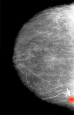
“In our trial, we used AI to identify screening examinations with a high risk of breast cancer, which underwent double reading by radiologists. The remaining examinations were classified as low risk and were read only by one radiologist. In the screen reading, radiologists used AI as detection support, in which it highlighted suspicious findings on the images”, says Kristina Lång, researcher and associate professor in diagnostic radiology at Lund University and consultant at Skåne University Hospital, who led the study.
The 80,033 women included in the safety analysis were randomly allocated into two groups: 40,003 women in the intervention group that underwent AI-supported screening and 40,030 in the control group that underwent standard double reading without AI support.
“We found that using AI resulted in the detection of 20 % (41) more cancers compared with standard screening, without affecting false positives. A false positive in screening occurs when a woman is recalled but cleared of suspicion of cancer after workup,” says Kristina Lång.
At the same time, the screen-reading workload for radiologists was reduced by 44 %. The number of screen readings with AI-supported screening was 46,345 compared with 83,231 with standard screening.
The time aspect is important. Kristina Lång explains that, on average, a radiologist reads 50 screening examinations per hour. The researchers estimated that it took approximately five months less of a radiologist’s time to read the roughly 40,000 screening examinations in the AI group.
“The study was conducted on a single site in a Swedish setting. We need to see whether these promising results hold up under other conditions, for example with other radiologists or other AI algorithms. There may be other ways to use AI in mammography screening, but these should preferably also need to be investigated in a prospective setting,” states Kristina Lång.
A total of 100,000 women have now been enrolled in the MASAI trial. The research team’s next step is to investigate which cancer types that were detected with and without AI support. The primary endpoint of the trial is interval-cancer rate. An interval cancer is a cancer diagnosed between screenings and generally have poorer prognosis than screen-detected cancers. The interval-cancer rate will be assessed after the 100,000 women in the trial have had at least a two-year follow up.
“Screening is complex. The balance between benefit and harm must always be taken into account. Just because a screening method finds more cancers does not necessarily mean it’s a better method. What’s important is to find a method that can identify clinically significant cancers at an early stage. However, this has to be balanced with the harm of false positives and the overdiagnosis of indolent cancers. The results from our first analysis shows that AI-supported screening is safe since the cancer detection rate did not decline despite a substantial reduction in the screen-reading workload. The planned analysis of interval cancers will show whether AI-supported screening also leads to a more accurate and effective screening programme,” says Kristina Lång.
For more information: https://www.lunduniversity.lu.se/
Related Breast Density Content:
VIDEO: FDA Update on the US National Density Reporting Standard - A Discussion on the Final Rule
One on One … with Wendie Berg, MD, PhD, FACR, FSBI
Task Force Issues New Draft Recommendation Statement on Screening for Breast Cancer
Creating Patient Equity: A Breast Density Legislative Update
AI Provides Accurate Breast Density Classification
VIDEO: The Impact of Breast Density Technology and Legislation
VIDEO: Personalized Breast Screening and Breast Density
VIDEO: Breast Cancer Awareness - Highlights of the NCoBC 2016 Conference
Fake News: Having Dense Breast Tissue is No Big Deal
The Manic World of Social Media and Breast Cancer: Gratitude and Grief
Related Breast Imaging Content:
Single vs. Multiple Architectural Distortion on Digital Breast Tomosynthesis
Today's Mammography Advancements
Digital Breast Tomosynthesis Spot Compression Clarifies Ambiguous Findings
AI DBT Impact on Mammography Post-breast Therapy
ImageCare Centers Unveils PINK Better Mammo Service Featuring Profound AI
Radiologist Fatigue, Experience Affect Breast Imaging Call Backs
Fewer Breast Cancer Cases Between Screening Rounds with 3-D Mammography
Study Finds Racial Disparities in Access to New Mammography Technology

 August 29, 2024


