Research image, courtesy of POSTECH.
March 10, 2023 — Breast cancer undisputedly has the highest incidence rate in female patients. Moreover, out of the six major cancers, it is the only one that has shown an increasing trend over the past 20 years. The chance of survival would be higher if breast cancer is detected and treated early. However, the survival rate drastically decreases to less than 75% after stage 3, which means early detection with regular medical check-ups is critical for reducing patient mortality. . Recently a research team at POSTECH developed an AI network system for ultrasonography to accurately detect and diagnose breast cancer.
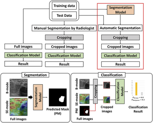
A team of researchers from POSTECH led by Professor Chulhong Kim (Department of Convergence IT Engineering, the Department of Electrical Engineering, and the Department of Mechanical Engineering), and Sampa Misra and Chiho Yoon (Department of Electrical Engineering) has developed a deep learning-based multimodal fusion network for segmentation and classification of breast cancers using B-mode and strain elastography ultrasound images. The findings from the study were published in Bioengineering & Translational Medicine.
Ultrasonography is one of the key medical imaging modalities for evaluating breast lesions. To distinguish benign from malignant lesions, computer-aided diagnosis (CAD) systems have offered radiologists a great deal of help by automatically segmenting and identifying features of lesions.
Here, the team presented deep learning (DL)-based methods to segment the lesions and then classify them as benign or malignant, using both B-mode and strain elastography (SE-mode) images. First of all, the team constructed a ‘weighted multimodal U-Net (W-MM-U-Net) model’ where the optimum weight is assigned on different imaging modalities to segment lesions, utilizing a weighted-skip connection method. Also, they proposed a ‘multimodal fusion framework (MFF)’ on cropped B-mode and SE-mode ultrasound (US) lesion images to classify benign and malignant lesions.
The MFF consists of an integrated feature network (IFN) and a decision network (DN). Unlike other recent fusion methods, the proposed MFF method can simultaneously learn complementary information from convolutional neural networks (CNN) that are trained with B-mode and SE-mode US images. The features of the CNN are ensembled using the multimodal EmbraceNet model, while DN classifies the images using those features.
The method predicted seven benign patients as being benign in three out of the five trials and six malignant patients as being malignant in five out of the five trials, according to the experimental results on the clinical data. This means the proposed method outperforms the conventional single and multimodal methods and would potentially enhance the classification accuracy of radiologists for breast cancer detection in US images.
Professor Chulhong Kim explained, “We were able to increase the accuracy of lesion segmentation by determining the importance of each input modal and automatically giving the proper weight.” He added, “We trained each deep learning model and the ensemble model at the same time to have a much better classification performance than the conventional single modal or other multimodal methods.”
This study was conducted with the support from the Ministry of Science and ICT, the Ministry of Education, and the Electronics and Telecommunications Research Institute (ETRI) of Korea.
For more information: https://international.postech.ac.kr/

 August 29, 2024


