April 22, 2015 — 3D Systems announced that a 20-month-old toddler is breathing and swallowing easier thanks to a team of cardiologists and cardiothoracic surgeons at Washington University School of Medicine in St. Louis. The team used a full-color 3-D printed replica of his heart to prepare for a delicate, 2.5-hour procedure at St. Louis Children's Hospital.
3DS features an end-to-end digital thread that integrates surgical simulation, training, planning and printing of anatomical models, surgical instruments and medical devices. The company has helped doctors in tens of thousands of complex medical cases to achieve better patient outcomes with faster surgeries.
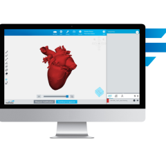
Life-size, realistic models make it simpler for patients and families to grasp the details of complex medical procedures, and they provide healthcare practitioners with invaluable preparation for their work in the operating room. In this particular case, the surgical team needed to relocate heart vessels that were squeezing and compressing the toddler's windpipe and esophagus, causing obstruction of the airway that resulted in difficulty breathing and swallowing. The printed model helped the team familiarize themselves with the unique vessel structure they would face in surgery, and they were also able to use it when discussing the condition with the patient's parents.
Shafkat Anwar, M.D., a member of the pediatric cardiology team at Washington University who worked with 3DS to develop the model heart for this particular surgical procedure, said, "With 3-D printing, we were able to print a replica of the patient's heart anatomy, developed from medical imaging scans, and use that model to get a handle on what surgeons would be faced with in the OR and to communicate with the patient's parents and other team members."
For more information: www.3dsystems.com


 April 18, 2025


