If you enjoy this content, please share it with a colleague
Siemens Healthineers
Siemens Healthineers brings together innovative imaging equipment, information technology, management consulting and services to help customers achieve tangible, sustainable clinical and financial outcomes. From imaging systems for diagnosis, to therapy equipment for treatment, to patient monitors to hearing instruments and beyond, Siemens innovations contribute to the health and well being of people across the globe, while improving operational efficiencies and optimizing workflow in hospitals, clinics, home health agencies, and doctors’ offices.
Videos
-
January 27, 2022
Cynthia McCollough, Ph.D., director of Mayo Clinic's CT Clinical Innovation Center, explains how photon-counting computed tomography (CT) detectors work and why it is a better technology over conventional CT systems. She helped Siemens develop the Naeotom Alpha, the first photo-counting CT system to be approved by the FDA in the fall of 2021. She spoke to ITN at the Radiological Society of North America (RSNA) 2021 annual meeting.
Read more about the first commercial photon-counting scanner
The device uses the emerging CT technology of photon-counting detectors, which can measure each individual X-ray photon that passes through a patient's body, as opposed to current systems which use detectors that measure the total energy contained in many X-rays at once. By "counting" each individual X-ray photon, more detailed information about the patient can be obtained and used to create images with less information that is not useful, such as image noise.
Current CT technology uses a two-step conversion process to convert X-ray photons into visible light using a scintillator layer in the detector. Then, photo diode light sensors turn the visible light into a digital signal. Due to this intermediate step, important information about the energy of the X-rays is lost and no longer available to aid in diagnosis. Also, contrast is reduced and images are not as clear.
Photon-counting detectors use a single step of direct conversion of X-rays into electrical current, and skips the step of converting X-rays into visible light. This allows the energy thresholds of each pulse to be collected and binned based on different kilovolt (kV) energy levels. This creates data to improve contrast and enable dual-energy, spectral imaging. The direct conversion also helps improve image quality without information loss. This improves image sharpness and contrast.
Photon-counting detectors have already been used for several years in high-energy physics and nuclear imaging. However, these previously generation photon-counting detectors could not be used with a clinical CT scanner because they could not keep up with the high higher rate of photons reaching the detector. The detector on the Naeotom Alpha was designed for this increased speed.
Related Photon-counting CT Content:
Mayo Clinic Begins Use of Third-Generation Photon-counting CT Clinical Research Detector
VIDEO: New Advances in CT Imaging Technology — Interview with Cynthia McCollough, Ph.D.
VIDEO: Photon Counting Detectors Will be the Next Major Advance in Computed Tomography — Interview with Todd Villines, M.D.
Key Trends in Cardiac CT at SCCT 2020
GE Healthcare Pioneers Photon Counting CT with Prismatic Sensors Acquisition
Top Trend Takeaways in Radiology From RSNA 2020
VIDEO: Advances in Cardiac CT Imaging — Interview with David Bluemke, M.D.
January 27, 2022Cynthia McCollough, Ph.D., director of Mayo Clinic's Computed Tomography (CT) Clinical Innovation Center, explains how CT dose tracking software works and offers advice to centers that record this patient level and device information. She spoke to ITN at the Radiological Society of North America (RSNA) 2021 annual meeting.
Dose tracking software allows hospitals and imaging centers to track what levels of radiation they are using by exam type protocol. It can show technologists who are using higher than required doses that may need additional ALARA training. The radiation dose tracking systems also can help track the amount of radiation a patient has received over time.
Related Radiation Dose Tracking Systems:
Disputed EHR Dose Levels Could Keep Patients From Necessary Imaging Exams
Medical Imaging Radiation Exposure in U.S. Dropped Over Past Decade
VIDEO: Radiation From Medical Imaging in U.S. Dropped Over Past Decade
The Basics of Radiation Dose Monitoring in Medical Imaging
VIDEO: Radiation Dose Monitoring in Medical Imaging — Interview with Mahadevappa Mahesh, Ph.D.
----
January 18, 2022Orlando Simonetti, Ph.D., professor, cardiovascular medicine, worked with Siemens to help develop a new, lower-field magnetic resonance imaging (MRI) system, the Magnetom Free.Max. It can scan patients that previously may have been contraindicated because of implantable medical devices. One of the first systems installed in the U.S. is at The Ohio State University Wexner Medical Center. It has a much lower magnetic field and a larger patient opening, removing barriers to MRI imaging for many patients.
Simonetti and his colleagues developed new techniques to boost the signal-to-noise ratio in MRI machines, which allowed the creation of a machine with a lower magnetic field strength that still enables high quality images.
The system gained FDA clearance in July 2021 and was featured by Siemens at the Radiological Society of North America (RSNA) 2021 meeting.
The interview and footage was provided by The Ohio University State University Wexner Medical Center.
Read more in the articles New FDA-approved MRI Expands Access to Life-saving Imaging and Ohio State Researchers Help Design New MRI, Expanding Access to Life-saving Imaging.
Related MRI Content:
Siemens Healthineers Announces First U.S. Installation of Magnetom Free.Max 80 cm MR Scanner
FDA Clears Siemens Healthineers Magnetom Free.Max 80 cm MR Scanner
Ohio State Researchers Help Design New MRI, Expanding Access to Life-saving Imaging
November 15, 2021Siemens and Philips demonstrated examples of new imaging software to convert MRI datasets into synthetic computed tomography (CT) datasets at the American Society of Radiation Oncology (ASTRO) 2021 meeting. The synthetic CT datasets can be used for radiotherapy treatment planning. This eliminates the need for a separate CT scan, reducing time and cost in patient care.
The technology uses an algorithm to convert the MRI dataset into a CT grayscale Hounsfield units. The Hounsfield units correlate with the densities of the various tissues and are used to calculate the doses required and beam routes needed in radiotherapy to treat a patient.
Photo Gallery of Technologies at ASTRO 2021
September 03, 2021ITN Editor Dave Fornell collected numerous examples of how PACS and enterprise imaging vendors are improving the speed and workflow of their systems during booth demonstrations at the 2021 Healthcare Information Management Systems Society (HIMSS). The 11 minute video condenses down the highlights of workflow efficiencies seen during two days o vendor booth tours.
There was a clear trend of many vendors moving to new platforms that leverage more modern cloud-platform interfaces. This enables faster study loading speeds over web connections. These platforms are also using deeper integration of third-party applications and artificial intelligence (AI) software that do not require separate logins or workflows. Read more about these key trends observed at HIMSS 2021.
Vendors also showed various ways they have speed up radiology workflows. These included easier to customize hanging protocols, automated fetching of prior exams, synchronizing views and scrolling between a current a prior exams, use of timeline views of patient priors and procedures to make it easier to find relevant images and reports, and integration of all types of images into one unified viewer.
Specific examples in this video include:
• Visage Imaging: Example of high speed cloud PACS access to 3D mammograms and and priors. This first video clip shows a demonstration of opening large datasets in a matter of a couple seconds over a network connection from a tethered cellphone.
• Visage Imaging: Ability to access multiple modalities on one PACS viewer
• GE Healthcare: Examples of fast access to priors and location on screen
• GE Healthcare: Example of deep integration of third-party AI software
• Siemens: Overview of its Lung AI Pathway Companion workflow
• Change Healthcare: Enabling fast ability to free rotate around lung anatomy rather than going slice by slice manually
• Change Healthcare: Color-coded bar shows loading progress of an image or data set
• Infinitt: Hanging protocol automation to find same view on prior and link for synchronized scrolling
• Infinitt: Use of timeline to get quick view of prior reports and images without needing to open whole exam
• Siemens: Example of deeper integration with third-party apps, in this case Epsilon strain echo analysis
• Fujifilm: Integrated advanced visualization in the radiology workflow for liver segmentation used for surgical or embolization planning
• Fujifilm: Example of life-like cinematic rendering of a CT scan offers new ways to view anatomy and explain it to a patient
• Visage Imaging: Example of enterprise platform able to bring in full original format advanced visualization reconstructed images on a single platform viewerRelated Medical Imaging IT Content From HIMSS 2021:
Advances in CVIS and Enterprise iImaging at HIMSS 21
Photo Gallery of New Technologies at HIMSS 2021
VIDEO: Importance of Body Part Labeling in Enterprise Imaging — Interview with Alex Towbin, M.D.
HIMSS 2021 Showed What to Expect From In-person Healthcare Conferences During the COVID Pandemic
VIDEO: Coordinating Followup for Radiology Incidental Findings — Interview with David Danhauer, M.D.
VIDEO: Cardiology AI Aggregates Patient Data and Enables Interactive Risk Assessments
VIDEO: Examples of COVID-19 CT Scan Analysis Software
August 31, 2021Several radiology IT vendors at 2021 Healthcare Information Management Systems Society (HIMSS) conference demonstrated computed tomography (CT) imaging advanced visualization software software to help automatically identify and quantify COVID-19 pneumonia in the lungs. These tools can help speed assessment of the lung involvement and serial tracking can be used to assess the patient's progress in the hospital and during long-COVID observation.
Examples of COVID analysis tool shown in this video include clips from booth tours at:
• Fujifilm
• Siemens Healthineers
• Canon (Vital)Canon received FDA clearance for its tool under and emergency use authorization (EUA).
Siemens said its tool was part of its lung analysis originally developed for cancer but modified and prioritized to aid in COVID assessments.
HIMSS Related Content:
Advances in CVIS and Enterprise iImaging at HIMSS 21
Photo Gallery of New Technologies at HIMSS 2021
VIDEO: Importance of Body Part Labeling in Enterprise Imaging — Interview with Alex Towbin, M.D.
VIDEO: Coordinating Followup for Radiology Incidental Findings — Interview with David Danhauer, M.D.
VIDEO: Cardiology AI Aggregates Patient Data and Enables Interactive Risk Assessments
VIDEO: Example of Epsilon Strain Imaging Deep Integration With Siemens CVIS
July 22, 2021This is an overview of trends and technologies in radiology artificial intelligence (AI) applications in 2021. Views were shared by 11 radiologists using AI and industry leaders, which include:
• Randy Hicks, M.D., MBA, radiologist and CEO of Reginal Medical Imaging (RMI), and an iCAD Profound AI user.
• Prof. Dr. Thomas Frauenfelder, University of Zurich, Institute for Diagnostic and Interventional Radiology, and Riverain AI user.
• Amy Patel, M.D., medical director of Liberty Hospital Women’s Imaging, assistant professor of radiology at UMKC, and user of Kios AI for breast ultrasound.
• Sham Sokka, Ph.D., vice president and head of innovation, precision diagnosis, Philips Healthcare.
• Ivo Dreisser, Siemens Healthineers, global marketing manager for the AI Rad Companion.
• Bill Lacey, vice president of medical informatics, Fujifilm Medical Systems USA.
• Karley Yoder, vice president and general manager, artificial intelligence, GE Healthcare.
• Georges Espada, head of Agfa Healthcare digital and computed radiography business unit.
• Pooja Rao, head of research and development and co-founder of Qure.ai.
• Jill Hamman, world-wide marketing manager at Carestream Health.
• Sebastian Nickel, Siemens Healthineers, global product manager for the AI Pathway Companion.
There has been a change in attitudes about AI on the expo floor at the Radiological Society of North America (RSNA) over the last two years. AI conversations were originally 101 level and discussed how AI technology could be trained to sort photos of dogs and cats. However, in 2020, with numerous FDA approvals for various AI applications, the conversations at RSNA, and industry wide, have shifted to that of accepting the validity of AI. Radiologists now want to discuss how a specific AI algorithm is going to help them save time, make more accurate diagnoses and make them more efficient.
With a higher level of maturity in AI and the technology seeing wider adoption, radiologists using it say AI gives them additional confidence in their diagnoses, and can even help readers who may not be deep experts in the exam type they are being asked to read.
With a myriad of new AI apps gaining regulatory approval from scores of imaging vendors, the biggest challenge for getting this technology into hospitals is an easy to integrate format. This has led to several vendors creating AI app stores. These allow AI apps to integrate easily into radiology workflows because the apps are already integrated as third-party software into a larger radiology vendors' IT platform.
There are now hundreds of AI applications that do a wide variety of analysis, from data analytics, image reconstruction, disease and anatomy identification, automating measurements and advanced visualization. The AI applications can be divided into 2 basic types — AI to improve workflow, and AI for clinical decision support, such as diagnostic aids.
On the workflow side, several vendors are leveraging AI to pull together all of a patients' information, prior exams and reports in one location and to digest the information so it is easier for the radiologist to consume. Often the AI pulls only data and priors that relate to a specific question being asked, based on the imaging protocol used for the exam. One example of this is the Siemens Healthineers AI Clinical Pathway and Siemens AI integrations with PACS to automate measurements and advanced visualization.
AI is also helping simplify complex tasks and help reduce the reading time on involved exams. One example of this is in 3-D breast tomosythesis with hundreds of images, which is rapidly replacing 2-D mammography, which only produces 4 images. Another example is automated image reconstruction algorithms to significantly reduce manual work. AI also is now being integrated directly into several vendors' imaging systems to speed workflow and improve image quality.
Vendors say AI is here to stay. They explain the future of AI will be automation to help improve image quality, simplify manual processes, improved diagnostic quality, new ways to analyze data, and workflow aids that operate in the background as part of a growing number of software solutions.
Several vendors at RSNA 2020 noted that AI's biggest impact in the coming years will be its ability to augment and speed the workflow for the small number of radiologists compared to the quickly growing elder patient populations worldwide. There also are applications in rural and developing countries were there are very low numbers of physicians or specialists.
Related AI in Medical Imaging Content:
AI Outperforms Humans in Creating Cancer Treatments, But Do Doctors Trust It?
VIDEO: Artificial Intelligence For MRI Helps Overcome Backlog of Exams Due to COVID
How AI is Helping the Fight Against Breast Cancer
VIDEO: Use of Artificial Intelligence in Nuclear Imaging
3 High-impact AI Market Trends in Radiology at RSNA 2019
Photo Gallery of New Imaging Technologies at RSNA 2019
VIDEO: Editors Choice of the Most Innovative New Radiology Technology at RSNA 2019
Study Reveals New Comprehensive AI Chest X-ray Solution Improves Radiologist Accuracy
VIDEO: Real-world Use of AI to Detect Hemorrhagic Stroke
The Radiology AI Evolution at RSNA 2019
Eliminating Bias from Healthcare AI Critical to Improve Health Equity
VIDEO: FDA Cleared Artificial Intelligence for Immediate Results of Head CT Scans
Building the Future of AI Through Data
Integrating Artificial Intelligence in Treatment Planning
Selecting an AI Marketplace for Radiology: Key Considerations for Healthcare Providers
Artificial Intelligence Improves Accuracy of Breast Ultrasound Diagnoses
Artificial Intelligence Greatly Speeds Radiation Therapy Treatment Planning
WEBINAR: Building the Bridge - How Imaging AI is Delivering Clinical Value Across the Care Continuum
AI in Medical Imaging Market to Reach $1.5B by 2024
VIDEO: AI-Assisted Automatic Ejection Fraction for Point-of-Care Ultrasound
5 Trends in Enterprise Imaging and PACS Systems
VIDEO: Artificial Intelligence to Automate CT Calcium Scoring and Radiomics
Scale AI in Imaging Now for the Post-COVID Era
VIDEO: Integrating Artificial Intelligence Into Radiologists Workflow
Northwestern Medicine Introduces Artificial Intelligence to Improve Ultrasound Imaging
Find more artificial intelligence news and video
August 07, 2019This is a quick walk around of the new Siemens Somatom Go.top cardiovascular edition compact computed tomography (CT) scanner on display at the Society Of Cardiovascular Computed Tomography (SCCT) 2019 meeting in July. It is aimed at cardiology office based imaging and was released this past spring at the American College of Cardiology (ACC) meeting.
The system has removable tablets on each side of the scanner where the tech can adjust the machine, review scout scans and trigger the scanner. The idea is to improve workflow and allow the tech to remain at the bedside longer to be with the patient, rather tucked away in a remote control room using an intercom.
The entire system is built into the gantry seen here, so there is no need for extra equipment in a closet, cabinet or server tower.
It comes in a 128 slice configuration with 4 cm of anatomical coverage per rotation.
It uses the Stellar detector and tin filtration to eliminate low energy photons and help lower dose. It can be programmed to aid workflow by automatically removing bone, create cured planar reconstructions, lung CAD and other post-processing features so more time can be spent on reading scans. The scanner also comes with a HeartFlow FFR-CT starter pack.
Find more information on this system in these related articles:
New Cardiovascular CT Technology Entering the Market
New Technology Highlights on the ACC 2019 Exhibit Floor
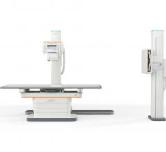
May 20, 2019This is a quick walk-around video showing the Siemens Healthineers Multix Impact digital radiography (DR) room X-ray system at RSNA 2018. The system offers an intuitive operating system to help improve productivity. The in-room touch user interface on the tube allows the technologist to remain at the patient’s side. And when unable to be at the patient’s side, the technologist is able to monitor and optimize positioning from the control room via the system’s patient positioning camera, potentially reducing repeat imaging and unnecessary patient dose. Lights at the top of the X-ray system automatically indicate it the patient anatomy is aligned properly with the exam type the technologist chose for optimal imaging to reduce the need for retakes.
The Multix Impact also offers an intuitive user interface and graphical organ program selection. The positioning guide display on both the in-room touch user interface and the workstation supports precise, consistent patient positioning. Advanced motorization and tracking reduce the physical exertion of technologists and help prevent repetitive stress injuries.
February 27, 2019This is a virtual heart with the same electrophysiology characteristics as the real patient unveiled by Siemens at the Healthcare Information Management and Systems Society (HIMSS) 2019 annual meeting in February. This "digital twin" technology is in development and will be able to create virtual, digital organs from a patient’s medical imnaging and other physiological data. In this case, the model was created using an ECG, MRI scan and other clinical data. It was shown as a way to help optimize cardiac resynchronization therapy (CRT) lead placement. CRT currently has a 30 percent nonresponder rate, which is mainly due to the placement of leads. This model allows virtual placement of the leads In various locations to test response prior to the implantation procedure. The green dot shows the location of the virtual lead. Siemens said the technology also might have applications for testing virtual ablations strategies to save procedure time when the patient is in the EP lab.
Read more about the digital twin technology.
Look through a photo gallery of other new technologies at HIMSS19.
Find news and videos from HIMSS 2019.
November 28, 2018This is an example of how artificial intelligence (AI) can help improve patient care by pulling together patient data from numerous sources and then select medical records that are specific to a patient’s diagnosis and treatment for a defined disease state. This is Siemens’ AI-Pathway Companion introduced at the Radiological Society Of North America (RSNA) 2018 meeting. In this examples. A prostate cancer patient has all their data on a single time line that can be accessed by single clicks on the points to open reports, images, procedures or labs.
At the end of the time line it integrates AI driven clinical decision support that recommends the next course of action based on clinical guidelines. The guidelines cited can also be opened for review by the clinician.
March 11, 2016Examples of technologies on the market and a discussion of what to look for in PACS and CVIS workflow efficiencies with Ascendian Healthcare Consultant Jef Williams. Editor Dave Fornell takes viewers on a tour of some of the key workflow improvements offered by health IT vendors in their software on the expo floor at Healthcare Information Management and Systems Society (HIMSS) 2016 meeting.
Related Content:
6 Key Health Information Technology Trends at HIMSS 2019
Technology Report: Enterprise Imaging
VIDEO: How to Build An Enterprise Imaging System
Enterprise Imaging 2018: Balancing Strategy and Technology
Three Resolutions Worth Keeping for a More Data-driven Radiology Practice
December 14, 2015Video discussion of new technology and trend highlights at the Radiological Society of North America (RSNA) 2015 meeting with ITN editor Dave Fornell and ITN contributing editor Greg Freiherr.
August 07, 2015ITN Editor Dave Fornell shares his choices for some of the most innovative new technology on the show floor at the 2015 AHRA meeting in Las Vegas.
May 26, 2015Detecting metastatic disease early is key. Sand Lake Imaging, Florida, provides great value to both patients and referrers by using syngo.via's efficient AV tools. See how!
April 28, 2015The new syngo.via from Siemens Healthcare supports oncological treatment decisions across modalities, therapies and departments to enable state-of-the-art cancer care, increased competitiveness, and high referrer and patient satisfaction. syngo.via can be used as a standalone device or together with a variety of syngo.via-based software options, which are medical devices in their own right. syngo.via VB10 and the syngo.via VB10 based software options are currently under development, and not for sale in the U.S., China and other countries. Due to regulatory reasons its future availability cannot be guaranteed. Please contact your local Siemens organization for further details.
October 17, 2014Siemens introduces True volume TEE transducer. This 3-D/4-D 90°x90° TEE solution enables clinically meaningful visualization of anatomy, volume color Doppler and function in one volume view, without compromises like stitching. Combined with eSieValves advanced analysis package, it offers automated modeling and quantification in seconds allowing cardiologists to remove the guesswork from valve sizing.
June 20, 2014DAIC Editor Dave Fornell shares his choices for the most innovative new technologies in nuclear imaging that were on display at the 2014 Society of Nuclear Medicine and Molecular Imaging (SNMMI) annual meeting.
June 13, 2014See how standardizing on Siemens' breast imaging solutions has helped Heritage Valley Health System optimize efficiency, clinical outcomes and patient experience.
May 15, 2014Improved patient comfort, more efficient workflow, and high-quality images at low dose are just some of the benefits realized in the radiology department at Philadelphia area's Riddle Memorial Hospital using Siemens fixed and mobile X-ray, and fluoroscopy solutions.
December 10, 2013Hear why Siemens SOMATOM Definition Edge is the CT your emergency department (ED) has been dreaming about from the leadership at Gwinett Medical Center. From physicians to the C-suite, see why the Edge is helping them meet their most demanding and time sensitive imaging needs with low-dose and high image quality.
RELATED CONTENT
News | PET-CTSiemens Healthineers’ new Biograph Vision positron emission tomography/computed tomography (PET/CT) system has been installed at the Hospital of the University of Pennsylvania (HUP) in Philadelphia – the first healthcare institution in the United States to install the technology.
South Texas Radiology Imaging Centers, San Antonio, recently became the first healthcare institution in the United States to install the Magnetom Sola 1.5 Tesla (1.5T), 70-cm magnetic resonance imaging (MRI) scanner from Siemens Healthineers. The Magnetom Sola features a new magnet design in addition to BioMatrix patient personalization technology.
Technology | AngiographyiSchemaView announced the release of RAPID Angio, a complete neuroimaging solution for the angiography suite that integrates iSchemaView’s RAPID software with syngo DynaCT Multiphase from Siemens Healthineers. The syngo DynaCT Multiphase is a three-dimensional image acquisition technique employing multiple rotations of a C-arm system to acquire a multi-phasic 3-D representation of the brain and its perfusion. This technology, when combined with the RAPID platform’s CTP product, delivers a powerful imaging solution to the angio suite for acute stroke patients.
Sponsored Content | Videos | Cardiac ImagingThis is a virtual heart with the same electrophysiology characteristics as the real patient unveiled by Siemens at the H ...
News | Artificial IntelligenceFebruary 13, 2019 — At the 2019 Healthcare Information and Management Systems Society (HIMSS) global conference and ...
News | Clinical Decision SupportSiemens Healthineers showcased the new planned artificial intelligence (AI)-based features with its mammography reading and reporting solution, syngo.Breast Care, at the 2018 Radiological Society of North America (RSNA) annual meeting, Nov. 25-30 in Chicago.1 These features are designed to provide physicians with interactive decision support.
Technology | Magnetic Resonance Imaging (MRI)January 30, 2019 — The U.S. Food and Drug Administration (FDA) has cleared the Magnetom Lumina 3 Tesla (3T) magnetic ...
News | Artificial IntelligenceSiemens Healthineers presented its first intelligent software assistant for radiology, the AI-Rad Companion Chest CT, at the 2018 Radiological Society of North America (RSNA) annual meeting, Nov. 25-30 in Chicago. The software brings artificial intelligence (AI) to computed tomography (CT). Using CT images of the chest, it can differentiate between various structures in that region of the body, highlighting them individually, and mark and measure potential abnormalities in the lungs, heart, aorta and coronary arteries. AI-Rad Companion Chest CT automatically translates its findings into structured reports.
Technology | Digital Radiography (DR)The U.S. Food and Drug Administration (FDA) has cleared the Multix Impact, an affordably priced, floor-mounted digital radiography (DR) system from Siemens Healthineers that expands access to high-quality imaging and enhances the patient experience.
News | Digital Radiography (DR)At the 104th Scientific Assembly and Annual Meeting of the Radiological Society of North America (RSNA), Nov. 25-30 in Chicago, Siemens Healthineers introduced the Multix Impact digital radiography (DR) system. The Multix Impact is an affordably priced, floor-mounted DR system that provides access to high-quality imaging technology. The intuitive operating system and versatile wireless detectors are designed to optimize clinical processes in radiography by improving productivity and enhancing the patient experience.
© Copyright Wainscot Media. All Rights Reserved.Subscribe NowE-newsletter Subscription form


 March 06, 2019


