It would be hard to argue that portable ultrasound isn’t among the most versatile imaging modalities in healthcare today. With improved imaging quality and ease of use, these units can routinely be found in departments across hospitals and clinics.
Portable ultrasound has arguably made the most significant impact on the emergency department (ED). In a relatively short period of time, ultrasound use in the ED has transformed many aspects of emergency medicine. The FAST exam (Focused Assessment with Sonography for Trauma) is a scan that helps determine internal bleeding and other injuries. This assessment has helped emergency medicine clinicians quickly determine the appropriate treatment.
“The FAST exam tells us within a minute or two whether there is life-threatening hemorrhage internally,” said Diku Mandavia, M.D., FACEP, FRCPC, an emergency medicine physician with Cedars-Sinai Medical Center and Los Angeles County Hospital. “That’s a really important data point for us. If someone arrives at time zero and I know within fifteen seconds if they have internal bleeding in their abdomen, that changes the direction of the resuscitation. I’ve been doing ultrasound for 16 years and practiced before ultrasound, and I saw the before and after. There were many patients who had delay in care and would, unfortunately, suffer consequences because of that. Now we have the opportunity to rapidly diagnose and get these patients to the operating room quicker, or to imaging, or to the ICU.”
But, emergency ultrasound is not limited to the FAST exam. Many newer procedures have developed in the emergency setting, according to Dr. Mandavia, that touch on both diagnostic and interventional areas of care.
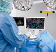
“There are newer features and techniques that are more specific to clinicians for guidance of procedures. When you talk about invasive lines into the central circulatory system or even lines into the peripheral system that may be difficult to achieve, ultrasound actually gives you point-of-care visualization,” Dr. Mandavia said. “It’s almost like cheating when doing a procedure. It’s so easy to put a central line in under ultrasound guidance. That is an evolving standard of care. That really helps patients from a safety perspective, so we don’t hit the carotid arteries or other serious targets.
“In [the ED], this is as much a physiological tool as it is an anatomic definition tool. Classically, ultrasound defines anatomy and pathology. You give ultrasound to a radiologist and they give you anatomy and pathology. You give that same technology to a clinician, and they add in critical physiology, which is amazing. We see patients who are in shock all the time. This is where the blood pressure is barely palpable; it could be in a peri-arrest situation and they may die imminently. The right interventions are critical when someone is that close to death. There are many different causes of shock and within a few moments, we can actually use ultrasound to define the cause of shock. That is essential to us and great for patients. This knowledge of the type of shock they’re in will help patient care. That’s an example of physiological assessment.”
Dr. Mandavia also sites vision loss as a condition that can be better evaluated in the ED, noting that ultrasound sees retina detachments better than fundoscopy.
Dr. Thomas Cook, M.D., program director, department of emergency medicine, Palmetto Health Richland, Columbia, SC, said ultrasound in the ED is the standard of care.
“As a residency director, I would say ultrasound in the emergency department is the standard of care,” Dr. Cook said. “The Residency Review Committee though Accreditation Council for Graduate Medical Education says you have to train emergency medical residents in ultrasound. Clearly for anyone who is up to date in the literature, it’s the standard of care.”
Purchasing Decisions
“I see a lot of manufacturers try to sell a small radiology system just to the emergency department,” Dr. Mandavia said. “That’s essentially not a good strategy and it never really works out well. We are primarily clinical physicians. We look after patients, and we use ultrasound as a technology in the emergency department to rapidly diagnose and treat patients. We want a system designed for simplicity of use to get to the right answers.”
In addition to high-diagnostic quality required to perform a variety of scans, durability and ease of use are key to effective portable ultrasound systems for the ED, according to Michael Lambert, M.D., of Advocate Christ Medical Center in Oak Lawn, IL. Clinicians stress that maneuverability is also among the main features that an ultrasound must have for the ED.
“Many EDs have tight spaces and weren’t designed ergonomically,” Dr. Cook said. “Laptop systems with a customized cart that is light, small and easy to move is usually ideal.”
Having a system with quick boot up capability, rapid acquisition of high-quality images and good presets are also key requirements, according to Dr. Mandavia. The rugged nature of the system also shouldn’t be forgotten.
“We recognize that the small radiology systems are good, but they weren’t built to withstand water spills or saline or blood or for things to bang into them,” Dr. Mandavia said. “You may have 100 staff in an emergency department, ranging from janitorial staff to physicians to nurses; things get moved around and bumped. You have fragile transducers sometimes. The design of a system is really important to us.”
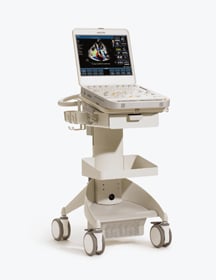
Cedars-Sinai Medical Center uses primarily SonoSite systems and one Aloka system, while the Los Angeles County Hospital uses ultrasounds from SonoSite, Aloka, Philips and GE. Palmetto Health Richland uses systems from GE, SonoSite and Ultrasonix, while Advocate Christ Medical Center uses models from SonoSite and Zonare. These hospitals all singled out SonoSite’s M-Turbo as one of the best portable units for ED use. In terms of future innovations, Dr. Lambert would like to see more user-friendly reporting packages and an easier process to export images to a DICOM reader.
“Our machines can do that now, but it seems like it could be less cumbersome,” he said.
Data management is another area that is ripe for improvement, according to Dr. Cook.
“I think you’ll see the manufacturers come out with some new data management solutions for [ED ultrasounds] in the next twelve months,” Dr. Cook said.
Cardiac Imaging
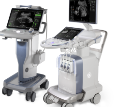
In August 2008, Philips Healthcare released its CX50 Compact Xtreme, designed to be a premium-level portable system for echocardiography. The system features the PureWave transducer, which aims to produce greater bandwidth and efficiency, yielding better imaging performance. This technology is designed to achieve greater penetration in patients who are traditionally difficult to image, such as obese patients or patients with emphysema. The system also features XRES adaptive imaging that reportedly reduces noise and artifact, producing greater clarity.
At Cleveland Clinic, the CX50 Compact Xtreme is used at the bedside, in the cath and EP lab, and in an outpatient setting.
“I feel comfortable using it in any environment,” said Richard Grimm, M.D., FACC, FASE, director of the echocardiography laboratory and a staff cardiologist in the section of cardiovascular imaging, department of cardiovascular medicine, at Cleveland Clinic Heart and Vascular Institute. “It could certainly be used as a stand-alone system in the outpatient clinic.”
“For standard exams, we use this system,” he added. “For a percentage of patients, we might go to a stand-alone high-end system for specialized 3D or different types of tissue Doppler capabilities, but that’s a minority of patients.”
Dr. Grimm said the CX50 Compact Xtreme compares very favorably to similar systems, citing ease of use and maneuverability, features that are advantageous for the clinic’s sonographers.

 July 19, 2024