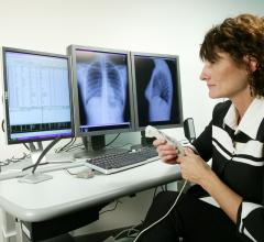
First used to treat brain tumors, radiosurgery has recently gone through a rapid evolution to include real-time image guidance and compact linear accelerators that can mount onto robotic arms.
During the last decade, radiosurgery has moved from solely an alternative to brain surgery to a viable non-invasive treatment for tumors anywhere in the body.
Improvements in image-guidance technology and robotics are the foundation on which a second-generation of radiosurgery systems has been built. These sophisticated radiosurgery systems have eliminated the need for patients to be fitted with a stereotactic frame and have expanded the use of radiosurgery beyond just intracranial applications. Now radiosurgery is capable of treating tumors throughout the body, including the head, neck, spine, lung, prostate, liver and pancreas.
Today’s radiosurgery systems use image guidance during the treatment process to determine the location of the tumor in real time. In the brain, the contours of the skull provide target reference coordinates, while elsewhere the body fiducials are typically implanted. However, new advanced algorithms are in use, which enable fiducial-free treatment of tumors both in the spine and the lung.
Continual image guidance during treatment
Continual, real-time image guidance is achieved by repeatedly determining the tumor location by using bony landmarks or the implanted fiducials. First the position of the tumor is determined prior to treatment by a computed tomography (CT) scan, which is often supplemented with an angiography or other type of functional imaging, such as a positron emission tomography (PET) scan or functional MRI (fMRI). These images are obtained while the patient is minimally restrained.
The supplemental images are then compared by the radiosurgery system’s computer with the images from the treatment planning CT. This enables the position of the tumor to be translated into the coordinate frame of the linear accelerator.
During the treatment process, real-time imaging is provided by X-ray devices. The radiosurgery system continually compares the X-ray images with the digitally reconstructed radiographs (DRRs) that were created during the treatment planning process. This comparison is what sets these second-generation radiosurgery systems apart because it enables the system and/or the clinician to check the tumor position and adjust the delivery of radiation accordingly if the tumor or patient moves.
These real-time adjustments are made possible by an interface between the treatment execution software and robotic devices that adjust the beam position during the treatment process. Currently there are two approaches to repositioning the patient during treatment. The first entails moving the table on which the patient rests during treatment. The second, more advanced method utilizes a robotic arm on which a lightweight linear accelerator is attached. In this scenario, the patient remains in the same position, but the robotic arm moves to change the beam target and trajectory.
Treating tumors that move
Using fiber optic sensing technology, the respiratory tracking technology is able to monitor and track a patient’s respiratory motions in real time. In conjunction with continual image guidance technology, respiratory tracking can correlate tumor motion with respiratory motion, dynamically directing the linear accelerator to deliver highly accurate radiation beams to moving tumors. Respiratory tracking technology constantly updates its correlation model with each new X-ray image, automatically correcting for any changes in the patient’s breathing patterns. This eliminates the need for complex gating or painful breath-holding techniques, allowing patients to breath normally during treatment.
Using a custom-designed vest with tracking markers, patient setup is simple, especially for multi-fraction treatments where the vest and markers rarely need to be adjusted on subsequent treatments.
A fiducial-free future
While fiducials still play a role in many of the extracranial radiosurgery treatments, technology is advancing to a point where even this minimally invasive process can be eliminated. Today, new advanced tracking algorithms have been developed that enable fiducial-free tracking of tumors in the spine and lung.
In the spine, the algorithm enables the real-time image guidance system to pick minute, discrete points on the vertebrae and track each of these points individually to determine patient or tumor movement during treatment.
For the lung, the first soft tissue tracking capabilities have been developed for continually monitoring the location of a lung tumor during treatment, even while the patient breathes normally. These use advanced, highly sophisticated image processing and image registration techniques to automatically lock onto the tumor directly, tracking it throughout the entire procedure.
Researchers are now exploring ways to eliminate the need for fiducials altogether, making radiosurgery completely non-invasive and expanding its uses to other indications, such as prostate and breast cancer. Achieving this goal will require further advancements in image guidance and treatment planning algorithms.
For instance, pre-operatively, there may be alternative ways to combine different imaging modalities, such as fusing PET with CTs and MRI, which would enable clinicians to more readily diagnose the tumor and determine its exact location.
Additionally, new advanced algorithms and other techniques may be developed to better compare the reference images.
Leksell’s prescient comment about new developments in physics and engineering still holds true today. Future developments, however, may not be making “radical changes in the old surgical handicraft.” Instead they will continue to shape the definition of radiosurgery and the approach in which cancer is treated.
Going mainstream
So what will it take for radiosurgery technology to become more of a mainstream option used by more than a handful of facilities?
Treatment experience over the past 40 years has established radiosurgery as an accepted option for the treatment of intracranial lesions. Over the past 10 years, Accuray Inc.’s CyberKnife Robotic Radiosurgery System has extended the application of radiosurgical principles beyond the brain to include extracranial targets such as spine, lung, liver, pancreas and prostate tumors.
Radiosurgery for the treatment of spinal tumors is now widely
accepted in the medical community worldwide. Acceptance of radiosurgery for the treatment of medically and some surgically inoperable lung tumors is also growing. This acceptance has led to the desire to compare radiosurgery to other proven surgical treatments, such as in the upcoming randomized study comparing CyberKnife radiosurgery to surgery for early-stage operable lung cancer patients. As more and more data are generated in treatment applications, such as prostate cancer and liver cancer, the acceptance of radiosurgery to treat these tumors and others will increase.
Costs to acquire and operate a system like CyberKnife also play a role. System and maintenance costs for the CyberKnife System are very similar to that of other radiosurgery-modified gantry systems. From an operational perspective, the daily QA staffing requirements for the CyberKnife System are notably less than that required for intensity-modulated radiation therapy (IMRT) systems.
Considering that the CyberKnife System is in the relatively early stages of its product lifecycle, its value is much greater when compared to other systems that are derived from technologies that have been on the market for 25-plus years and may be nearing the end of their lifecycle.
Many facilities have found that use of the CyberKnife System has improved their overall patient throughput because an entire radiosurgery treatment can be completed in 1 to 5 days, as compared to the weeks and months required to complete treatment with conventional radiation delivery technology. The CyberKnife System’s accuracy and tissue-sparing abilities enable it to deliver much higher doses of radiation in 1 to 5 treatment sessions.
What is more interesting, however, is that many facilities that have installed the CyberKnife System have experienced an overall increase in radiation oncology referrals to the facility. This is because patients who are appropriate candidates for radiosurgery can be treated in addition to patients whose disease is more amenable to treatment with conventional radiotherapy. The ability of the CyberKnife System to be complementary to existing conventional treatments expands the range of patients to whom a site can offer treatment.
While the field of radiation oncology is constantly innovating and developing new methods for compensating for target motion and improving methods of radiation dose delivery, the
CyberKnife continuously tracks target motion and can automatically correct for that motion and ensure that the high doses of radiation reach the target accurately and safely through its Synchrony Respiratory Tracking System. The CyberKnife continues to improve its methods of tracking and target identification, as exemplified by the introduction of the completely non-invasive real-time lung tumor tracking system, Xsight Lung, at the 2006 ASTRO meeting.
Radiosurgery and the CyberKnife System are about accuracy and not simply delivering high doses. To best treat patients it is essential to spare as much normal tissue as possible and to ensure the tumor receives the full dose. Conventional radiation delivery systems and “part-time” radiosurgical systems are constrained by the fixed rotations of their gantry systems and the inability to automatically correct for tumor motion. Most of these systems rely solely on pretreatment imaging to target tumors, and do not continuously assess tumor location during the treatment. This has the potential to drastically reduce their ability to accurately deliver treatments and maximally spare sensitive normal tissue.
Euan S. Thomson, Ph.D., is president and CEO, Sunnyvale, CA-based Accuray Inc., which develops and markets the CyberKnife Robotic Radiosurgery System. Thomson has served as Accuray’s CEO since March 2002, and as president since October 2002. From March 1999 to February 2002, Thomson served during various periods as president, CEO and a member of the board of directors of Photoelectron Corp., a publicly held medical device company. Prior to joining Photoelectron, he held various positions as a medical physicist within the United Kingdom National Health Service and worked as a consultant for medical device companies, including Varian Oncology Systems and Radionics Inc. Thomson holds a B.S. in Physics, an M.S. in Radiation Physics and a Ph.D. in Physics, with an emphasis on stereotactic brain radiotherapy, each from the University of London.


 July 28, 2017


