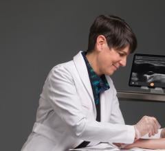
Cardiac ultrasound technology has advanced to keep up with several trends. These include improved workflow for greater efficiency, expanded use of qualification metrics, expanded use of 3-D echo to speed exam times and improve operator reproducibility, and expanded use of 3-D transesophageal echo (TEE) to aid guidance in the growing area of transcatheter structural heart procedures. Here are a few examples of how the newest technology is addressing these trends.
Improving Reproducibility
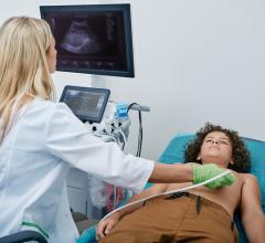
A major issue in ultrasound is the variation between operators, which leads to different measurements for the same anatomy in the same patient based on operator experience and technique. Operator variability may also limit the use of ultrasound as a cardiac triage tool as it expands into point of care.
3-D echo may solve part of this issue by enabling acquisition of volume datasets, rather than very specific slices of the anatomy. A 3-D volume acquisition allows a cardiologist reviewing the exam to select the optimal slices during post-processing, instead of relying on the echo tech to get the specific 2-D views. Some high-end ultrasound systems already extract the ideal views automatically.
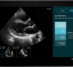
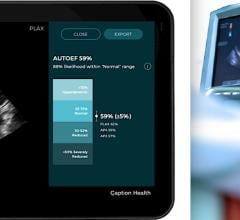
In addition, most high-end echo systems now automatically quantify measures such as ejection fraction, left ventricular mass and strain values. An example of this is GE Healthcare’s Automated Function Imaging (AFI) software on several of its systems, which automates 2-D speckle tracking to measure in real time the deformation (strain) of the myocardial wall. The algorithm tracks the wall motion and calculates the percentage of lengthening or shortening in a set of three longitudinal 2-D image planes (apical long, two chamber and four chamber) and displays the results for each plane. It then combines the results of all three planes in a single bull’s-eye summary, which presents the analysis for each segment along with a global peak strain for the left ventricle.
In 2013, Philips Heathcare introduced its new Epiq ultrasound platform. The system was built from the ground up with new technology and software, including use of Philips’ Anatomical Intelligence software, which is designed to help operators get the optimal views needed for specific exams and measurements. Anatomical Intelligence has artificial intelligence components that on later editions will eventually help identify what anatomy and organs are being imaged and from what direction, which can help guide less experienced operators to get perfect exam views. Only about 25 percent of the Anatomical Intelligence software is currently deployed in the first version of Epiq’s cardiology release. Much of it offers ideal illustrations to show the operator how images should appear for specific views. This and other workflow improvements for mitral valve evaluations can cut these exams from 10 minutes down to two to three minutes, Philips said.
Epiq’s nSIGHT technology is a new imaging architecture that provides highly detailed images and temporal resolution, allowing for clinicians to see new levels of tissue uniformity and penetrate at higher frequencies for improved views of typically difficult-to-image patients.
Workflow and Image Quality Improvements
At the 2014 American College of Cardiology (ACC) meeting in March, Esaote premiered its new CrystaLine imaging technology that improves ultrasound’s diagnostic efficiency by enhancing image clarity and providing clinicians the ability to personalize image quality for each patient. The technology contains several workflow enhancements to help clinicians efficiently and effectively manage their increased workload with the expected influx of new patients in the coming years.
XView+ and CPI are two CrystaLine features that enhance diagnostic confidence and improve clinical workflow by increasing image clarity across patient populations, from obese to pediatric. XView+ is a unique speckle reduction algorithm that provides multiple levels of noise reduction so sonographers can personalize image quality by “dialing in” the right amount for each patient. CPI improves image quality at depth so sonographers can more effectively image structures deeper within the body of large and difficult-to-scan patients. Images for all patients now yield better anatomical definition throughout extended fields of view, leading to a more confident interpretation of image data.
Toshiba America Medical Systems launched its new cardiovascular echo platforms in 2013, the Aplio 500 and Aplio 300 CV (cardiovascular) systems. Both are for premium 2-D cardiac exams and feature Toshiba’s 2-D Wall Motion Tracking technology, which provides visualization and quantitative analysis of myocardial wall motion with accuracy and reproducibility. The systems offer on-board cardiac quantification measurements in all directions (radial, circumferential, 2-D rotation and longitudinal). Additional cardiac-specific technologies include tissue enhancement, advanced dynamic flow, lateral gain controls, tissue Doppler, stress echo, Flex-M mode and auto IMT. Both systems are designed for ease of use with ergonomics in mind and a smaller footprint.
True 3-D Echo Visualization
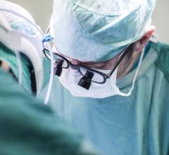
At the 2013 Transcatheter Cardiovascular Therapuetics (TCT) meeting, GE unveiled its XDclear technology, which increases penetration by 2-4 cm and improves high-definition resolution throughout the image. It also allows true 3-D visualization for echo images created by the Vivid E9 ultrasound system. It uses 3-D echo datasets to create a 3-D image on screen. When the operator wears a pair of PolarVision glasses, the image becomes true 3-D, making it much easier to differentiate depth. To further enhance this feature, GE also created Depth Illumination, which aids depth perception with the addition of synthetic shadowing. It makes anatomy look more like a real surgical view, as if the patient’s anatomy is exposed and seen under surgical lighting. GE demonstrated these combined technologies from a recorded MitraClip procedure on 3-D/4-D TEE. It offered a very clear image of the device’s position, which is often not very clear on traditional 2-D or 3-D TEE.
TEE Advances
With the growing number of new transcatheter procedures for mitral valve repair, transcatheter aortic valve repair (TAVR), left atrial appendage (LAA) closure and closure of atrial septal defects (ASDs) and ventricular septal defects (VSDs), 3-D TEE is becoming an essential tool for procedural navigation.
In 2013, Philips updated its CX50 xMatrix portable ultrasound system to interface with its live 3-D TEE. The smaller footprint is expected to make it more competitive in the cath lab and hybrid OR environments. Its architecture also supports Philips’ EchoNavigator, which aligns TEE views with live fluoroscopy.
GE’s Vivid E9 Breakthrough 2012 (BT12) cardiac ultrasound system includes a 3-D/4-D TEE probe, offering tools to improve workflow through simplified image acquisition. It is designed for intuitive navigation and yet easy-to-use quantification. The TEE probe allows 4-D dataset acquisitions. The probe includes triplane imaging, 4-D views and high frame rates. It enables one-button acquisition of mitral valve images, rather than using the track ball and a combination of buttons to slice the image into different views. In bi-plane mode, it has the ability to tilt and rotate at the same time. The system also offers laser lines, a new tool to help understand the relationship between 2-D slices and 4-D views, helping to visualize the linkage between 2-D and 4-D.
Siemens fourSight TEE View enables 3-D acquisition, reconstruction and display at the point of care. Both 2-D and 3-D assessment can be combined with the use of a V5Ms transducer. Reconstruction can be done immediately or at a later date.
 March 06, 2024