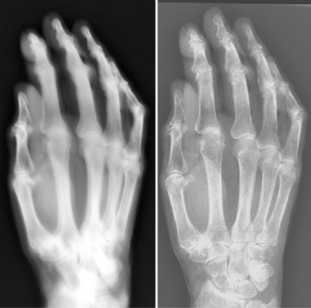
A comparison of an X-ray of a hand using 1896 technology (left) and a modern digital radiography X-ray system.
Researchers recently tested first-generation X-ray equipment from 1896 and found it produced radiation doses and exposure times that were vastly higher than those of today’s systems, according to a study published online and in the May print edition of Radiology.
“To my knowledge, nobody had ever done systematic measurements on this equipment, since by the time one had the tools, these systems had been replaced by more sophisticated ones,” said the study’s lead author, Gerrit J. Kemerink, Ph.D., from Maastricht University Medical Center in the Netherlands.
Wilhelm Roentgen reported his discovery of X-rays on Dec. 28, 1895. A few weeks later, H.J. Hoffmans, a physicist and high school director in Maastricht, the Netherlands, and L.T. van Kleef, M.D., director of a local hospital, performed anatomical imaging experiments with an X-ray system built from equipment at Hoffmans’ high school. Key elements of the system included a high-voltage transformer and a glass bulb with metal electrodes at each end.
Technology advanced rapidly, and the setup used by Hoffmans and Dr. van Kleef soon became obsolete. Eventually, the equipment ended up collecting dust in a Maastricht warehouse. A year ago, Jos M.A. van Engelshoven, M.D., Ph.D., former radiology head at the Maastricht University Medical Center, retrieved the equipment, most of which was still in working order, for a television program on the history of healthcare in the region. Kemerink then decided to analyze the setup in more detail.
The Maastricht researchers repeated some of the first imaging exams, using the equipment to image a hand specimen from a body that had been donated to science.
“We sometimes worked in a fully dark room that had black walls, with the only light coming from the flashing tube and from discharges in the spark gap,” Kemerink said. “Together with the irregular buzz of the interrupter and the crackling sound of the discharges, this created a very special, kind of ghostly, ambiance.”
The researchers compared the radiation dose, X-ray beam properties and electrical characteristics of the 1896 system with those from a modern X-ray system. Using the same exposure conditions used in 1896, the estimated skin dose needed to image the hand was nearly 1,500 times greater on the first-generation system than on the modern system — 74 milligrays (mGy) and 0.05 mGy, respectively. Corresponding exposure times were 90 minutes for the old system and 21 milliseconds for the modern system.
Pinhole images showed the X-rays originated from an extended area of the glass wall in the system’s construction, causing image blurring. Still, the 114-year-old system produced what Kemerink described as surprisingly good images in which anatomical details were clearly visible.
The high radiation doses and long exposures times of early X-ray equipment caused significant health problems for the technology’s pioneers. Adverse effects, such as eye complaints, skin burns and loss of hair, were reported within weeks of Roentgen’s discovery.
“Many operators of the early X-ray systems experienced severe damage to hands over time, often necessitating amputations or other surgery,” Kemerink said.
X-ray technology improved rapidly in the 20th century, with significantly lower radiation dose and exposure time and improved image quality, making it a convenient and safe imaging modality and an invaluable diagnostic tool.
Reference: Collaborating with Drs. Kemerink and van Engelshoven were Martijn Kemerink, Ph.D., Tom J. Dierichs, B.S., Julien Dierichs, B.S., Hubert J.M. Huynen, and Joachim E. Wildberger, M.D., Ph.D. “Characteristics of a First-Generation X-Ray System.” Radiology. March, 2011
For more information: http://radiology.rsna.org

 July 18, 2024