With the development of 3D-CRT, IMRT, stereotactic approaches and other advanced forms of radiation therapy, imaging has become increasingly important in the field of radiation oncology. These more precise treatment modalities make it possible for us to plan and deliver very conformal radiation doses, in keeping with the main goal of radiation therapy, which is to maximize the dose to the tumor volume and minimize the dose to normal tissues.
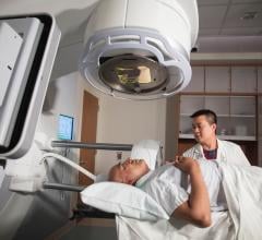
Image courtesy of Varian Medical Systems. Varian's Trilogy linear accelerator is equipped with the On-Board Imager device.
However, the ability to shape dose distributions so precisely resulted in a number of new challenges. To aim a very conformal beam at a tumor, the clinician must know exactly where it is during treatment, or risk delivering too little dose to some part of the target or too much to normal healthy tissue. The variability of tumor position over a five-to six-week course of treatment makes this problematic. Even with the use of immobilization devices and alignment of the equipment with external marks on the patient’s skin, there is day-to-day variation in patient and tumor position. During treatment, there is the problem of intra-fraction motion due primarily to respiration.
Traditionally, we have not had the means for exactly localizing the target site each day, and so the standard of care was to treat a larger volume around the tumor, while holding doses low. This was a trade-off. Although research has shown that tumor control is improved with higher doses, it was important not to exceed normal tissue tolerances.
The advent of image-guided radiation therapy (IGRT) is changing the equation. Very simply, IGRT involves the use of imaging tools to localize the area being treated, as well as verify how treatment was delivered to the tumor. IGRT tools and techniques help clinicians deal with both patient positioning uncertainties and with tumor motion due to respiration.
There are a number of new IGRT tools available today, and many are still evolving. There are room and gantry-mounted kV imaging systems, optical tracking devices with 3D ultrasound reconstruction technology and respiratory gating systems used to synchronize imaging and dose delivery with a patient’s breathing cycle.
The ideal IGRT system is one that can produce different kinds of images just prior to treatment and then help clinicians make use of those images in a clinically practical way. Clinicians will require online visualization of bony anatomy, soft tissue — with or without implanted markers — and real-time tumor motion. Sophisticated software then compares the images with reference images from earlier treatment planning, calculates the “delta,” or how the patient must be moved in 3D so that the tumor lines up with the radiation beams, and instructs the treatment couch accordingly.
IGRT has recently opened the door to true four dimensional (4D) radiation treatment. In addition to dealing with the three dimensions of space, radiation therapy techniques also deal more effectively with the problem of tumor motion in time — the fourth dimension. Using 4D imaging, 4D simulation, 4D treatment planning, 4D treatment delivery and 4D verification, clinicians will be able to precisely determine the margin around the tumor and program the technology to deliver treatment that adapts to the tumor motion, keeping that margin constant. Ultimately, that is the path that IGRT has forged. For some tumor sites, such as the brain or the head and neck, IGRT will help shrink margins by eliminating patient set-up uncertainties. For tumors in the chest or abdomen where respiratory motion has a very big effect, IGRT presents a solution toward overcoming this problem.


 June 07, 2022


