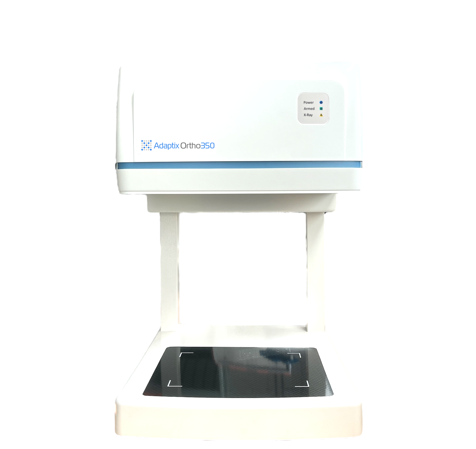
Adaptix Ortho350, uses 3D X-Ray technology and an image reconstruction algorithm to provide 3D orthopedic imaging at a cost and radiation dose similar to existing 2D X-Ray systems.
As resource pressures continue in Emergency Departments, patients with relatively minor injuries are increasingly redirected to Minor Injuries Units or triaged by specialist nursing teams with the power to request and review imaging services.
While this has real benefits in terms of reducing waiting times and focusing emergency resources on the most seriously ill or injured patients, the number of "missed fracture” claims facing the National Health Service (NHS) has increased notably over recent years, costing the health service over £1.1million between 2015 and 2018 (NHS Resolution Report, 2022).
The reasons for misdiagnosis naturally vary from case to case, but the Resolution Report highlights two consistent failings across England: diagnostic error, particularly early incorrect diagnoses of soft-tissue injuries; and issues with requests for imaging, reporting, interpretation and follow up — including failure to complete further imaging such as CT or MRI that is indicated in national guidance.
Challenges
Perhaps most worryingly, given the increasing responsibility being placed on specialist nursing teams to triage patients, the majority of errors highlighted in the report were due to clinical judgment mistakes, with incorrect initial diagnoses meaning crucial X-rays were not requested in nearly half of cases reported. The role of advanced nurse practitioners and radiologists for initial triage and diagnosis has many advantages for patient flow, but the checks and balances that should be in place to ensure any errors are picked up quickly are not always realized due to resource pressures.
At the simplest level, if the triage team misdiagnoses a soft-tissue injury when a fracture is present, imaging may not be requested, and the patient may be sent home with inappropriate soft-tissue care advice. Similarly, if X-ray imaging is requested, fractures — particularly those that are difficult to spot, such as scaphoid fractures, may not be picked up by less specialist teams, leading to similar outcomes.
In both cases, specialty reviews of all cases should take place within set time frames. For example, NICE guideline NG38 states: “A radiologist, radiographer or other trained reporter should deliver a definitive written report of Emergency Department X-rays of suspected fractures before the patient is discharged from the emergency department” (Section 1.1.9). This recommendation is reiterated in the Royal College of Emergency Medicine's Management of Radiology Results in the Emergency Department, which states: “All results of radiological investigations performed in the Emergency Department must be reviewed by a clinician, taking clinical scenario into account, and necessary actions taken.”
However, where staffing levels and resource pressures are not sufficient to support these safeguards — as is the case in many units across the country — mistakes will occur, meaning injuries will be missed and patients will be placed at risk of suboptimal outcomes.
In a similar vein, 3D imaging technologies such as MRI and CT have become the “gold standard” for many orthopedic diagnoses, but limited resources and long imaging waiting lists mean most U.K. clinicians rely on X-rays, which may not show all fractures in the first instance.
NICE's own Resource Impact Report acknowledges there are resource implications of implementing its guidelines, stating that “the use of MRI/CT as first line imaging in suspected spinal injuries will place demands on an already limited resource” (Section 3.2.2). It also states that “there will be additional cost implications to provide this service out of hours” (Section 3.3.5) and that, “while national guidelines support immediate reporting, in practice this process is often delayed.”
In most cases, this will simply mean a few additional days of pain and delayed treatment for patients, but for others, getting it wrong can have life-changing consequences.
New Imaging Technology
So how do we square the circle of increasing patient demand and over-stretched resources to provide the best service possible for patients, and without breaking ED budgets?
One answer lies in investment in new imaging technologies that support non-radiologists to diagnose and screen minor injuries more effectively.
Both NICE and Royal College of Emergency Medicine guidelines highlight the importance of “right imaging, right place, right time” diagnostics and greater use of new imaging technologies offers real potential to improve patient outcomes cost-effectively.
For example, one such innovation, Adaptix Ortho350, uses Adaptix’ 3D X-Ray technology and an image reconstruction algorithm (developed in partnership with the University of Oxford) to provide 3D orthopedic imaging at a cost and radiation dose similar to existing 2D X-Ray systems. It covers many different angles, allowing depth information to be derived and presented in DICOM format but, unlike other Digital Tomosynthesis (DT) systems that can experience difficulties localizing some structures due to limited depth resolution, the Adaptix system "sweeps" in two dimensions, allowing enhanced ‘Z’ resolution, improving image resolution.
This delivers several benefits for clinicians, including enhanced diagnostic confidence and a reduced need to refer patients for expensive MRI or CT imaging.
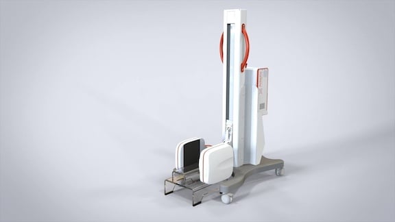
This next-generation imaging technology also means 3D imaging can be obtained more easily at the point of patient care — be that in the waiting room, by the patient’s bedside or even in an ambulance or community setting — reducing the need to move patients around the hospital and even preventing unnecessary admissions.
With quicker image acquisition times — up to 30 second to take the image and provide full image reconstruction — clinicians can review images in almost real time, allowing faster diagnosis and treatment, greatly increased diagnostic confidence, and reduced waiting times.
This accelerated workflow not only reduces costs but also improves patient care, reducing the risk of misdiagnosis by making 3D imaging more accessible and reducing the need to move patients around the hospital, which can potentially make injuries worse and increase cross-infection risks.
Several of these imaging technologies have already been proven in other, highly-regulated industries and their deployment around the healthcare estate could be one of the most cost-effective investments our healthcare services could make.
Better imaging, available at the point of care, speeding up and increasing accuracy of diagnosis, reducing waiting lists, saving unnecessary and expensive imaging and radiation exposure, as well as reducing the huge financial cost of misdiagnoses? All at the cost of existing 2D X-Ray technology? What’s not to like?

Siân Phillips, MD, is a consultant radiologist in the National Health Service and chief medical officer at Adaptix, Oxford, England, United Kingdom.

 December 03, 2025


