The tried and true old workhorse of imaging modalities, particularly for women’s healthcare, has demonstrated its longevity but it’s also evolving to address future needs in the face of newer and more advanced imaging modalities.
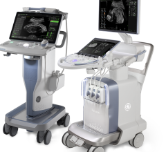
That includes adopting at least one computed tomography characteristic and 4D capabilities. Healthcare facilities also see considerable growth in compact, hand-held models, as well as ergonomic design in mobile, cart-based models.
So what might the future hold for ultrasound technology?
Outpatient Care Technology sought answers and insights from some of the leading ultrasound equipment manufacturers on how their products are developing and serving outpatient care facilities.
What are some of the more recent noteworthy improvements in ultrasound units and why do they matter clinically and operationally?
Omar Ishrak, president and CEO, clinical systems business unit, GE Healthcare
Three key innovations are volume imaging, ultrasound architecture evolving from hardware-based to software-based architecture and miniaturization. First, volume imaging dramatically improves visualization of anatomy, giving clinicians information in 3D. The real-time nature of ultrasound creates 4D. This gives clinicians confidence in understanding anatomy and function. For example, our cardiology customers talk about the added clinical confidence 4D imaging provides by visualizing the heart in 3D and in real-time.
Volume imaging also improves standardization and productivity, greatly reducing the need for re-scans and increasing the ability to see more patients. Second, software-based architecture has enabled the rapid development of software-based algorithms to better view images. A software-based architecture improves the ease of use by networking images and enabling wireless connections to PACS, EMR and data management systems. Now ultrasound can ride the same technology curve as the computer and telecommunication industries for the miniaturization of hardware. Third, compact ultrasound systems the size of laptop computers create portability, allowing clinicians to take this to new markets for new uses and reach more patients.
Al Lawson, vice president, strategic marketing, Resonant Medical Inc.
For customers of Resonant Medical, clinical and operational improvements in ultrasound units translate into efficiently converting ultrasound data into precise patient positioning and anatomical-structure targeting information used throughout the radiotherapy planning and treatment delivery process. Within radiation oncology, the essential workflow elements must all converge to provide the most accurate daily delivery of the prescribed therapeutic dose of radiation. Accurate delivery of the daily treatment depends on reproducing the conditions (3D or 4D) represented by the treatment plan through the precise alignment of patient to the planned isocenter created within a treatment plan to the isocenter of the linear accelerator.
Intensity Modulated Radiation Therapy permits the radiation oncologist to prescribe sharp dose gradients to organs and adjacent/contiguous volumes based on their risk for disease involvement or tolerance to radiation damage. Patient anatomy is not static and therefore rapid and precise 3D definition and alignment of the patient setup according to the plan are critical. The radiation oncologist’s prescription has been meticulously derived from multimodality imaging sources (CT, PET-CT, MR and ultrasound) fused into a common data set and used as a reference for that patient within the treatment room.
In the future, the user will require greater automation to rapidly make a determination of the patient’s current position and align them to the intended coordinates for treatment. Decision support software (i.e., is the patient correctly positioned) will provide the user an opportunity to compare these to the original plan and then consider when to revise or adapt the original radiotherapy plan. Clearly, to make these decisions the user will require auto-segmentation (structure rendering of the live time ultrasound imaging), auto-fusion of the current image to the multimodality reference image (which includes ultrasound information), and clear guidelines derived from the radiation oncologist’s prescription (e.g., an estimate of the 3D coverage accuracy for today’s session).
Tim Dugan, senior vice president, U.S. sales and global marketing, SonoSite Inc.
The advent of faster and more powerful Digital Signal Processors (DSPs) and the continued advances of Application Specific Integrated Circuit (ASIC) technology have really propelled ultrasound performance into new levels of performance. The large, cart-based systems have leveraged this for 3D technology and other research applications. At SonoSite, we are most excited about how these technologies have accelerated the levels of performance now available in the portable point-of-care markets. In the past few months alone, we have added a proprietary image processing technology called SonoMB, which has dramatically improved the contrast resolution and overall image presentation on our flagship MicroMaxx product. The really exciting thing about this release is that the flexibility already designed into the product using these DSP and ASIC components has allowed us to implement this capability in a ‘software’ only upgrade.
Ultrasound saw a rapid improvement in cost-performance when the ‘digital’ beamformer era started in the 1990s. We are now at a point where the hardware is become fast and flexible enough that major advances in image quality performance can be made through software upgrades. To our customers, this means better performance and more value, both now and in the future.
Gordon Parhar, director, ultrasound business unit, Toshiba America Medical Systems
Most noteworthy is the hand-carried ultrasound (HCU) market, which is driven by two technology trends – advanced PC-based architecture and rapid miniaturization. Growing from a $5 million market in 1999 to more than $200 million in 2006, these high-performance, portable and affordable systems are accelerating the proliferation of ultrasound imaging to medical specialties other than cardiology, radiology and OB/GYN. This is driving adoption of ultrasound imaging by new user groups like surgeons, anesthesiologists and emergency physicians, enabling increased patient access and improved improves quality of care.
How do you make ultrasound units even more user-friendly without
compromising effectiveness?
ISHRAK: More sophisticated algorithms, advances in software platforms and imbedded protocols could further automate image optimization. New applications that are probe-specific for a clinical market could be another ease-of-use advancement. Other advances in transducer technology could be lighter probes with wider bandwidth, making them easier to use with better image quality.
LAWSON: Resonant Medical spends considerable R&D to simplify the user interaction and optimize the timely acquisition and 3D rendering of the B-mode ultrasound image. Our goal is to convey real high-quality information to the user rather than merely data. The data may be useful to others for post treatment analyses, but the user in the treatment room needs information for decision support and assure them they are accurately delivering the therapeutic dose as intended. Radiation oncology, as with all outpatient departments, must provide their services in a timely and efficient manner, while never minimizing the accuracy demanded. The delivery of a therapeutic dose of radiation is irreversible, and therefore the patient’s anatomy must be positioned correctly every session or we fail to give the patient the best chances for cure or possibly increase the risk for unintended side effects. Radiation oncology information systems offer the opportunity to verify and record the parameters of the how the linear accelerator was positioned, but the live time ultrasound addresses where the anatomical based positioning to the accelerator’s isocenter.
DUGAN: This issue has always been a struggle for the high-end cart-based systems. Oftentimes, the need to provide a lot of functionality conflicts with the need to provide fast and easy-to-use systems. In the point-of-care space, the name of the game is efficiency and effectiveness. We really need to focus on providing the best image on a wide variety of patients in the shortest amount of time possible.
At SonoSite, we have a relatively large department in R&D that is dedicated to Human Factors Engineering. This group focuses on finding the right balance of ease-of-use and flexibility. We have also spent countless hours optimizing the system controls so various imaging parameters are automatically adjusted and optimized in the background with commonly used controls such as depth. Another feature we are proud of is the new ‘Auto Gain’ control, which automatically adjusts gain and other internal parameters based on the actual image processed by to the system. These advances have helped give our ultrasound systems a ‘bigger sweet spot,’ allowing the clinician to focus on the patient instead of on our system.
PARHAR: To improve effectiveness, new technologies must facilitate automated operation so results can be reproduced by different operators. The accuracy of the sonographic exam is almost exclusively dependent on the skill and experience of the sonographer. Eliminating operator dependency is the key challenge for ultrasound. Automated 3D/4D (volumetric imaging) significantly eliminates operator dependency, standardizing the exam and improving reproducibility.
What do you foresee as the next big ultrasound development to expand functionality or improve performance in medical imaging? How do you envision next-generation ultrasound units functioning or performing differently? What additional capabilities and features will be available to improve patient care in the outpatient setting?
ISHRAK: The next significant development in ultrasound would be the growing integration of ultrasound as a guidance tool for other procedures. Using ultrasound to visualize procedures such as nerve blocks for regional anesthesia will help drive safety and quality of care. The next-generation units could be more application-specific for clinicians in emerging markets. As we continue the path of designing for each user, ultrasound might not look like the ultrasound systems we see today. The continuation of miniaturization and advancements in software-based architecture will fuel these improvements in patient care.
LAWSON: The radiation oncology community wants technologies that assure the precise delivery of the intended therapeutic dose of radiation. We believe that 3D ultrasound deployed throughout the treatment process (simulation, planning and treatment room) offers a cost effective and efficient solution for all patients positioning for a variety of diseases. Currently, daily ultrasound imaging for positioning prostate cancer patients prior to treatment is common. However, other anatomical sites also require conformal delivery techniques for optimal care, e.g., conformal treatment techniques for partial breast irradiation. Inherent in their conformal nature, restricting dose to the tissue at risk is critical. Increasing the radiation dose to either the remainder of involved breast or the contralateral breast is not compatible with the intent to restrict the unintended dose. Three-dimensional ultrasound breast imaging offers essential imaging information without the use of ionizing radiation and would seem like an obvious choice. For this to be useful probe pressure to obtain the 3D ultrasound must be non-perturbing to the natural anatomy, and the technique to produce the image must be rapid and quickly allow the user to position the patient accurately. Good images should be produced rapidly by the user without user interaction with the image. Once optimal settings are derived within a clinic they should be maintained for that clinical situation by patient.
DUGAN: As ultrasound devices get smaller, there will be opportunities for dedicated devices to assist in performing procedures. We have already seen a rapidly growing demand for the use of ultrasound in guiding line placements and regional nerve block procedures. We see the same happening in the future for guiding radiation therapy. Size and ease-of-use will be critical in taking ultrasound from the dedicated diagnostic modality that it once was, to a useful tool for the clinicians to help them do their jobs better.
In after-loading applications, because technology is allowing ultrasound to get smaller, we see a big increase in the use of point-of-care to evaluate patients either at the bed-side or at outpatient settings. Key elements to accelerating this adoption are uncompromised image quality and seamless connectivity to the hospital’s IT infrastructure.
PARHAR: As in MRI, CT and conventional X-ray, the use of contrast media could change the way ultrasound is performed, opening new and unique diagnostic opportunities. Contrast agents, once approved by the FDA for radiology applications, will improve the image quality either by decreasing the reflectivity of the undesired interfaces or by increasing the backscattered echoes from the desired regions. Contrast harmonic imaging, CHI, is a Toshiba-exclusive technique, opening the possibility of measuring blood perfusion or capillary blood flow.
Contrast imaging, along with miniaturization and volume imaging will open new doors for ultrasound. Having a HCU at the patient’s bedside to monitor the location of the drug delivery agents with the precision of volume ultrasound will improve the quality of care.
[Next-generation ultrasound units] will be faster and smaller with better image quality. They will be easier to operate with more automation and more quantitative data to assist physicians with diagnosis. Additionally, multimodality image fusion will be possible with ultrasound and CT/MRI images fused together to provide more conclusive diagnosis.
Additional features could include ultrasound-guided drug delivery in oncology once developers can control the cavitation effect produced by ultrasound beams that allow drugs to pass through the protective membranes of cells. It may also include using ultrasound more frequently for [radiofrequency] ablation since ultrasound is a cost-efficient way to guide the ablation probe and monitor the procedure.
Feature | October 14, 2007 | Rick Dana Barlow
Integration, miniaturization drive modality’s progress


 March 19, 2025
