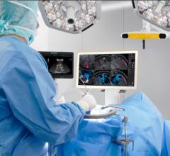
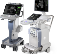
The Epiq ultrasound system from Philips Healthcare features Anatomical Intelligence software in combination with a new imaging architecture called nSIGHT. The system combines high-quality imaging with anatomic structural models as guides to make exams more reproducible and easier to perform.
As a less expensive, radiation-free form of medical imaging, ultrasound is finding increased utility in many areas of healthcare. The technology’s evolving flexibility and portability make it an attractive option for doctors looking to triage patients in an emergency room, perform rapid cardiology examinations or offer supplemental imaging for women who have already undergone screening mammograms. Its use is rapidly expanding at the point of care in hospitals in more than 20 specialties, according to a December 2013 report in the journal of the World Heart Federation, Global Heart.1
Some of the imaging modality’s advancements in recent years include image quality improvements and the emergence of tissue elastography and volumetric ultrasound. These improvements and more are pushing ultrasound into the forefront for care in a variety of settings.
Improving Skill Sets With
Technology, Training
It is known that successful use of ultrasound often depends on the machine operator. Operator variability can affect clinical confidence in an ultrasound imaging exam when factors such as patient anatomy or an unfamiliar vendor, system or transducer are in play. One example of an advancement that can address this is Philips Healthcare’s new Epiq ultrasound system, which features a proprietary imaging technology that comes in combination with Philips’ Anatomical Intelligence. Epiq received 510(k) clearance from the U.S. Food and Drug Administration (FDA) in August 2013 and launched at the 2013 European Society of Cardiology (ESC) Congress in Amsterdam last summer.
Epiq’s nSIGHT technology is a new imaging architecture that provides highly detailed ultrasound images and temporal resolution, allowing for clinicians to see new levels of tissue uniformity and penetrate at higher frequencies for improved views of typically difficult-to-image patients. In addition, the Anatomical Intelligence software and its database of anatomic structural models provide users with advanced organ modeling, imaging slicing and quantification, making exams more reproducible and easier to perform. Epiq is the first Philips system to have Anatomical Intelligence built in, recognizing a growing need for ultrasound examinations to be reproducible without relying on a specialized operator.
Another trend that aims to improve the use of ultrasound is its utility as an educational tool for future physicians. In September 2012, first-year medical students at Mount Sinai School of Medicine were issued GE Healthcare’s Vscan pocket ultrasound devices during their white coat ceremony.2 At a size only slightly larger than that of a smartphone, the Vscan device (first introduced in 2010) provides an immediate, noninvasive method to image key areas of the body. The distribution was part of a research study to determine if hand-held imaging technologies could significantly contribute to medical education at all levels of training.
The study, led by Bret Nelson, M.D., associate professor in the department of emergency medicine at Mount Sinai, was reviewed in a paper titled “Point-of-Care Cardiac Ultrasound: Feasibility of Performance by Noncardiologists” in the December 2013 Global Heart. The clinic-based study demonstrated that the first-year medical students were able to detect pathology in 75 percent of patients with known cardiac disease, whereas board-certified cardiologists using stethoscopes could detect only 49 percent.1Nelson and co-author Amy Sanghvi, M.D., instructor in emergency medicine at Mount Sinai, also described in the paper the growing use of focused ultrasound in many areas outside of cardiology, where it is traditionally used. “Increased training by clinicians across many specialties, coupled with technology improvements yielding lower cost and better quality studies, have contributed to this trend,” they said in a press release.
In another paper in the December 2013 Global Heart journal, authors led by J. Christian Fox, M.D., professor of clinical emergency medicine and director of instructional ultrasound, University of California Irvine School of Medicine, advocated for the inclusion of ultrasound in medical curriculum to help doctors become more adept at using the modality for detecting potentially life-threatening conditions.3The authors also urged the earlier inclusion of ultrasound in training in order “to fully realize its benefits as early as possible,” referring to a study by Kobal et al4 that demonstrates the potential benefits of extending ultrasound learning in medical education.
Increasing Portability Leads to Increased Applications
Technological advancements in ultrasound are helping pave the way for more doctors to arm themselves with the imaging tool when approaching a variety of cases at the point of care. At the 2013 annual meeting of the Radiological Society of North America (RSNA), several vendors showcased their latest ultrasound systems, including pocket devices to compete with GE’s Vscan, which was the first to enter the market.
New vendors in portable ultrasound include Konica Minolta, which launched the Sonimage P3 hand-held ultrasound device to bring its imaging capabilities to the patient or bedside, helping clinicians visually assess a patient immediately when determining a diagnosis. The personal ultrasound device is easy to use and can easily fit into a lab coat pocket or be worn like a stethoscope. Weighing less than 14 ounces, the device offers B-mode, M-mode and Doppler mode, as well as simple-to-access presets. It can be used as a standalone ultrasound system or plugged into a Windows-based PC, laptop or tablet for the flexibility of a larger display.
Also on display at RSNA 2013 was Samsung and its UGEO lineup of ultrasound systems to complement existing Samsung Medison systems. These included the high-end UGEO HM70A, a new hand-held ultrasound system that can be deployed across specialties such as radiology, cardiology and OB/GYN, and UGEO PT60A, a tablet-based ultrasound system that is compact and easy-to-use for point-of-care applications.
Samsung also showed its work-in-progress UGEO WS80A ultrasound system, a device geared specifically toward clinicians in OB/GYN. The UGEO WS80A features “5-D” imaging that automatically extracts relevant anatomical views from the acquired volumetric data for a study, and could have potential application in cardiology. It includes a 21.5-inch LED screen with 10.1-inch touch panel and breast elastography capabilities to simplify the distinguishing of benign masses from malignant ones through acquiring the strain ratio between the target and reference area. This device was first showcased at the 23rd International Society of Ultrasound in Obstetrics and Gynecology (ISUOG) World Congress, held last fall in Sydney, Australia.
Another new point-of-care system, the X-Porte ultrasound kiosk from SonoSite (now part of Fujifilm), received 501(k) clearance from the FDA in November 2013. It utilizes SonoSite’s proprietary Extreme Definition Imaging (XDI) technology that was developed following 35 years of applied ultrasound research. The X-Porte offers visual learning guides, integrating high-resolution imaging with 3-D animations to enable any member of a healthcare team to maximize use of the ultrasound technology. The system is available in a stationary or detachable-use model.
Lasting Trend
The advent of more pocket-sized, portable ultrasound devices and improved imaging technologies in areas such as elastography and volumetric imaging make ultrasound an attractive alternative to other modalities such as computed tomography (CT) and magnetic resonance imaging (MRI). Its availability and accessibility also make ultrasound a unique tool for physicians who need immediate imaging to assess a patient, whether at the bedside, in a clinic or in remote areas. These latest advancements in ultrasound will continue to trend in an era where doctors and hospitals are continuously looking to cut costs and radiation dose while still maintaining positive outcomes for patients.
References
1. Nelson BP, Sanghvi A. “Point-of-Care Cardiac Ultrasound: Feasibility of Performance by Noncardiologists.” Global Heart. 2013 Dec;8(4):293-97.
2. “Mount Sinai School of Medicine Issues Pocket Ultrasound to Students.” Diagnostic and Interventional Cardiology. 2012 Nov/Dec;52(6):34.
3. Christian Fox J, Marino H, Fischetti C. “Differential Diagnosis of Cardiovascular Symptoms: Setting the Expectations for the Ultrasound Examination and Medical Education.” Global Heart. 2013 Dec;8(4):289-92.
4. Kobal SL, Trento L, Baharami S, et al. “Comparison of effectiveness of hand-carried ultrasound to bedside cardiovascular physical examination.” Am J Cardiol. 2005;96:1002–06.


 July 19, 2024
