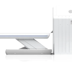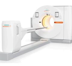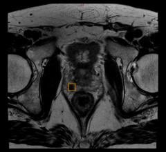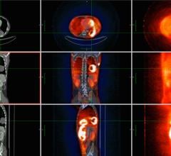
The Gemini TF PET/CT applies time-of-flight technology for improved event localization and image quality.
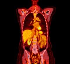
Traditionally, CT has been used for staging and radiation treatment planning. However, it is not always sufficient for tumor localization and characterization and requires a follow-up PET scan. As recent studies have shown that concurrent PET/CT improves accuracy for radiation therapy planning over separate PET and CT scans, there is a growing trend in oncology toward the adoption of hybrid PET/CT modalities for multiple image functions – diagnosis, staging and treatment planning.
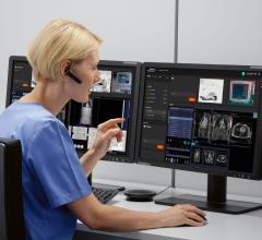
Over the years, has become more sophisticated in co-registering images acquired separately and is expected to continue to play an important role in multimodality imaging in the coming years. Agfa HealthCare is currently developing its works-in-progress IMPAX Registration and Fusion, a solution designed to allow for
single modality registration for follow-up studies (CT/CT, MR/MR), mult-modality fusion for anatomical studies (CT/MR) or a combination of anatomical and physiological studies (tracers in PET images). The solution will be ideally suited for noninvasive diagnosis and surgical treatment planning.
Moving forward, the next step is to acquire back-to-back images using hybrid scanners, such as PET/CT. Historically in radiation oncology, CT images alone have been used for IMRT (Intensity Modulated Radiation Therapy) treatment planning. However, clinicians are increasingly fusing PET with CT images to perform PET/CT-guided IMRT. Since IMRT depends to a large degree on accurate delineation of targets and critical structures, it is very important that the co-registration of fused PET and CT scans for IMRT are extremely accurate. The same holds true for PET-guided radiation therapy (PGRT), which involves fusing diagnostic PET images with treatment planning CT images taken in the radiation oncology department, often from exams performed on different days.
While clinicians have suspected that using a hybrid PET/CT system might be more advantageous than PET and CT images acquired separately and later fused, researchers at the Washington University School of Medicine in St. Louis, MO, put the question to the test. The researchers did find that the structures identified on the PET images demonstrated better correlation to patient anatomy on the CT taken at the time of PET than to patient anatomy on the traditional separate CT. What surprised researchers was that the CT image acquired at the time as the PET was not any more accurate than the CT taken at a separate time. But what produced better results was taking the PET and CT images consecutively, in the same treatment
session, with the patient in the same position and with minimal time interval.
As images improve, so should treatment, and high-quality images have been driving progress in PET-guided radiation therapy. For example, tumor localization has reached a new level at University of Washington, Seattle, where 4-D whole-body respiratory gated PET/CT imaging is now routine. Using the GE Discovery STE PET/CT, clinicians are able to scan for simultaneous acquisition of whole-body respiratory gated PET and a list mode coincidence file. As a result, “4-D PET has been acquired as part of the clinical routine and is currently conducted on all whole-body PET/CT exams,” indicated Paul Kinahan, associate professor of Bioengineering, University of Washington. In addition, respiratory motion of the lung, liver and heart can be captured and assessed across the entire patient FOV.
Sound image resolution is the objective behind the design of Philips Medical Systems’ Gemini TF PET/CT, a three-dimensional (3-D) PET scanner with a 16-slice Brilliance CT scanner. Its time-of-flight (TOF) technology, TruFlight, measures the time difference between the detection of two coincident gamma rays, then uses this time difference to more accurately localize the origin of the annihilation. Improved event localization reduces noise in image data, resulting in higher image quality.
As the hybrid modality continues to improve upon image quality in radiation therapy, PET/CT’s applications are expected to expand beyond oncology, to play an important role in cardiovascular disease, measuring myocardial viability, and for diagnosis of neurological diseases such as Parkinson’s, Alzheimer’s and epilepsy.
Experts anticipate that the trend toward using hybrid PET/CT in oncology and beyond will continue, as long as better images lead to better treatment.


 July 02, 2024
