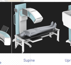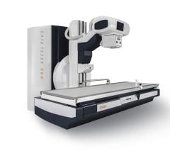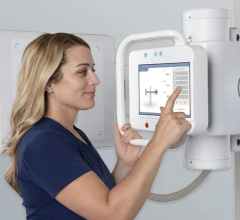
The Carestream DRX Revolution DR system.
Since its introduction to the marketplace 20-plus years ago, digital radiography (DR) has been making an impact.
A white paper by Frost & Sullivan from June 2009 estimated that information from nearly 70 percent of all imaging procedures was in digital format at that time.1 More recently, a report issued by Millennium Research Group in November 2010 discusses how DR is growing and notes that expected expansion of the total U.S. X-ray system market through 2015 will be propelled in part “by the adoption of more expensive digital flat-panel detector systems.”2 In addition, “Practice Guideline for Digital Radiography,” developed jointly by the American College of Radiology (ACR), American Association of Physicists in Medicine (AAPM) and Society for Imaging Informatics in Medicine (SIIM), talks about the “paradigm shift” from analog screen-film to digital imaging.3
The widespread adoption of DR is natural, given the substantial benefits it offers over film. An online study module on DR by Sprawls Educational Foundation outlines a number of DR’s advantages.4 Foremost is the ability of digital images to be processed. There are various forms of post-processing that can be used to enhance contrast and the visibility of some details, such as computer-aided detection (CAD). In addition, the viewer can zoom, compare multiple images and perform a variety of analytical functions.
Also, digital radiographic receptors have a wide dynamic range, while that of film is somewhat limited. This means a good image contrast can be formed over a wide range of exposures. Potentially, comparable image quality can be obtained by digital radiography at a lower dose than film.
However, DR’s ability to compensate for underexposures and overexposures can have a downside, despite its advantages. Sprawls’ discussion points out two potential problems: “Even though images with good contrast can be produced with relatively low exposures, they will have a high level of quantum noise…the level of image (quantum) noise depends on the exposure to the receptor. When a low exposure is used, the result can be excessive image noise.
“The other problem is that excessively high and unnecessary exposures can be used to form images. While these images will have good quality (low noise), there will be unnecessary exposure to the patient. This problem does not exist with film radiography, because the increased exposure will result in a visibly overexposed film.”
The joint Practice Guideline includes a similar caveat: “On the one hand, retakes due to inappropriate exposure are reduced, but overexposures can easily be hidden, as there is no direct feedback given by the image presentation.”
Regardless of that limitation, there are even more benefits to note. Digital images are available immediately, and they can be stored and retrieved rapidly. They can be copied without loss of image quality. And they offer much more accessibility for remote locations, whether the locations are other areas of the imaging facility itself or across the world.
Using DR can also lower some of the costs associated with processing, managing and storing films. For example, DR images require much less physical storage space than film. Also, as the Frost & Sullivan report indicates, the time to do a digital X-ray is less than two minutes per exam, while older analog systems can take 15 minutes. This increases a facility’s throughput and efficiency.
The Frost & Sullivan report also says the switch to electronic medical records (EMRs) is encouraging a conversion from analog to digital: “Digital radiography enables a patient’s radiographic images to be tied to his electronic medical record, allowing them all to be stored together, to be read quickly and efficiently when recalled.”
There are numerous vendors offering DR systems, including several who recently introduced wireless models. Specifications for a number of systems are available on the following pages. An expanded chart is available online at www.itnonline.net.
References:
1 “Digital Imaging Technologies Reduce Costs.” Frost & Sullivan White Paper, June 2009, carestream.com/medit-resources.html
2 Introduction to “U.S. Markets for X-Ray Systems 2011.” Millennium Research Group, November 2010, mrg.net/Products-and-Services/Syndicated-Report.aspx?r=RPUS50XR10
3 “ACR-AAPM-SIIM Practice Guideline for Digital Radiography.” 2007, www.acr.org
4 “Digital Radiography” study module. Sprawls Educational Foundation, www.sprawls.org/resources/DIGRAD/module.htm

 June 21, 2024

