Radiation therapy has played an important role in the treatment of cancer for more than a century. Used typically as a curative treatment either alone or in conjunction with surgery and/or chemotherapy, the aim of radiation therapy has always been to eradicate a patient’s cancer.
Technology has improved and the delivery of radiation has become more precise, and radiation oncologists are now able to deliver higher doses of radiation more safely. This has given radiation an even bigger role in cancer care.
Radiation therapy has a wide range of uses including as an adjuvant therapy, in which radiation is used after surgery to sterilize a surgical site of residual cancer cells. In neoadjuvant therapy, radiation is used prior to surgery to reduce the size of a tumor, sterilize the area around a tumor and improve surgical outcomes. Radiation therapy can also be used in the palliative setting to alleviate symptoms associated with tumors — such as pain and bleeding — in order to improve a patient’s quality of life.
Types of Radiation Therapy Treatments
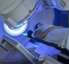
Radiation therapy is typically broken down into two subcategories: external beam radiation and internal radiation, or brachytherapy. During brachytherapy, radioactive seeds or pellets are implanted close to or within a tumor site. This can be done either during an open surgery, or outside of a surgical setting using needles, catheters or tubes. External radiation involves the use of radiation machines to deliver X-ray, electron or proton radiation to a tumor.
There have been many advances that have revolutionized the way external beam radiation is delivered.
Progression of X-ray Therapy
X-ray therapy has greatly improved in the last few decades. Unlike early cobalt radiation that used low-energy X-rays, today, radiation oncologists utilize high-energy, megavoltage X-rays that can penetrate more deeply and minimize irradiation of the skin. According to American Society of Radiologic Technologists (ASRT) Vice President Sandra Hayden, M.A., R.T.(T)., multileaf collimators (MLC) replaced traditional shielding with blocks. In addition, MLC allowed radiation therapy professionals to provide intensity modulated radiation therapy (IMRT) — little leaflets that can move in and out of the beam to modulate the fluence pattern of the beam. “IMRT is a very sophisticated type of X-ray treatment, where instead of relying on just a few static beams to deliver radiation, multiple actively modulated beams are utilized,” said Henry Tsai, M.D., ProCure Proton Therapy Center, New Jersey. He said that in IMRT, each of the beams is actively modulated during the delivery so that the shape of the distribution of radiation can be more tightly wrapped around the tumor, minimizing exposure of the surrounding tissue.
The use of MLC has continued to increase in radiation X-ray therapy. In February 2013, the UC Davis Comprehensive Cancer Center was one of the first sites in North America to install an MLC system on its linear accelerators. The center reported in a press release that the MLC system allowed radiation oncologists to customize the therapeutic beams to conform to a tumor’s shape and size.
Advancements of Imaging in Radiation Therapy
In addition to the advancement of IMRT, Hayden said that image-guided radiation therapy (IGRT) is becoming a mainstay. IGRT utilizes enhancements or attachments in addition to the delivery component of radiation. For example, cone beam computed tomography (CBCT)/kilovoltage (kV) imaging panels mounted onto treatment units allow radiation therapists the ability to acquire imaging information prior to and during treatment. This imaging information is used to guide radiation beams and treat tumors.
Tsai said that combining high-resolution imaging studies with radiation delivery allows for better outcomes. “The better we can define where the tumor is, the better ability we have to improve the therapeutic ratio, minimizing the exposure to healthy tissues that can occur with radiation treatment.” Hayden echoed this statement saying, “This technology allows radiation therapists and radiation oncologists to have an even better visualization of structures as well as the tumor, to deliver radiation therapy more precisely than ever before.”
Research continues to push the limits of IGRT. This past May the Netherlands Cancer Institute-Antoni van Leeuwenhoek Hospital, Amsterdam, the Netherlands, joined the University of Texas MD Anderson Cancer Center, Houston, and the University Medical Center Utrecht, the Netherlands, as part of a research consortium that will help to develop an image-guided treatment combining radiation therapy with magnetic resonance imaging (MRI), creating an MRI-guided radiation therapy system. “MRI is the gold standard modality for imaging soft tissues, and MR imaging of the cancer during radiation therapy could provide the healthcare team the ability to optimize treatment while reducing toxicity,” said Steven J. Frank, M.D., associate professor of radiation oncology, director of advanced technologies, MD Anderson, in a press release. Hayden added that MRI “is being utilized for its superior quality and will continue to enable us to target the tumor and limit radiation dose to surrounding areas.”
Functional and metabolic imaging is also undergoing further research. “Functional and metabolic imaging will help us target the radiation delivery by enabling us to see not just where the tumor is, but also where the metabolically active parts of the tumor are,” Tsai said. He said that by also potentially allowing physicians to view the hypoxic areas of tumors — areas that may be more resistant to treatment and therefore might require higher doses of radiation or radiosensitizers — functional and metabolic imaging will help guide physicians in where they need to intensify cancer treatment.
Advancements in Proton Therapy
According to Tsai, the next phase of technology that is becoming more prevalent is proton therapy, the most advanced form of radiation available. Proton beams are much more precise. “Pencil beam scanning, also known as spot scanning, has the ability to treat complex tumors while avoiding healthy tissues and critical structures,” Hayden said.
Proton therapy’s reduction of normal tissue exposure can be credited to physicians’ ability to control how far proton beams penetrate into the body. “Unlike an X-ray, which enters the body and exits out of the other side, exposing a lot of the normal tissues to unnecessary radiation, a proton beam can be controlled. We can decide how far we want that beam to enter the body. The beam delivers its energy at the specified depth and then stops moving,” Tsai said. This method eliminates exit radiation, giving physicians the ability to increase radiation dose and more powerfully target tumors. “We can take advantage of that proton property to deliver more radiation to tumors while minimizing the amount of radiation that is given to the surrounding healthy tissues,” he continued. Hayden said that because radiation is confined to the tumor, side effects generally common after radiation therapy are reduced with proton therapy.
Due to its success and benefits, proton therapy is gaining widespread use in different areas of cancer treatment and research. According to clinical study results presented at the American Society for Radiation Oncology’s (ASTRO) 55th annual meeting, proton therapy has proven to be a cost-effective treatment for pediatric brain tumor patients. In June, Loma Linda University Medical Center, Loma Linda, Calif., announced that it had openings in eight clinical trials for patients with breast, prostate, liver and pancreas cancers — all involving the use of proton therapy.
As research rapidly sheds light on the benefits of this technique, academic centers are building proton therapy centers around the United States. Emory Healthcare and the Winship Cancer Institute’s Emory Proton Therapy Center in Atlanta and the Texas Center for Proton Therapy in North Texas are both scheduled to open in 2016. According to a report by the market research firm RNCOS, there may be as many as 27 proton centers in the United States by 2017.
Conventional Surgery vs. Radiosurgery
Due to the increased precision of radiation beams, one trend that will likely see an increase is the decision of patients to forego surgery and opt instead for radiosurgery or stereotactic ablative radiation therapy. In radiosurgery and stereotactic ablative radiation therapy, high doses of radiation are targeted at small, well-defined tumors of the brain, lung, liver and spine, with the goal of killing the cancer, while also minimizing exposure to surrounding healthy organs, Hayden said. These techniques offer a noninvasive, nonsurgical option for tumors. “Although in some cases radiation is used as an adjunct to surgery, we maybe seeing more and more patients turning to radiosurgery as an option for certain types of tumors,” Tsai said.
Additional Advancements
According to American Cancer Society data, breast cancer patients in particular are living longer due in part to advancements in early detection. Physicians have begun to incorporate patients’ breathing patterns into simulation. The gating/deep inspiratory breath hold (DIBH) technique requires that patients hold a deep breath for a short period of time in order to allow the radiation oncology team to target the radiation exactly where it needs to go. “Utilizing gating, there is less possibility of long-term cardiac toxicity,” Hayden said.
Healthcare and Radiation Therapy
As healthcare encourages the use of more cost-effective solutions, making hospitals and healthcare centers increasingly cost-conscious, many would think that radiation therapy would take a hit due to its association with higher costs. However, Tsai does not see this as a likely scenario. In many cases, technological improvements have actually resulted in more cost-efficient delivery of radiation therapy. “Although IMRT machines and proton machines may be considered pricey technologies, as we improve them and understand how we can make treatment systems more compact and less expensive, ultimately we can deliver the same, sophisticated radiation treatments but not incur the large cost associated with it,” Tsai said.
Improving Individualized Treatment
The expanding and maturing population in the United States could possibly lead to more cancer diagnoses in the future, making cancer a more prevalent problem. Radiation therapy will continue to be a necessary and vital part of cancer treatment.
Cancer research is working toward the ability to predict what mode of treatment will offer a specific patient the best outcome. Tsai said, “For some cancer treatments or biologic agents/chemotherapies, we know that a certain percentage of patients may not respond to the treatment, but the question is, which patients are those?”
Cancer research is helping physicians better figure this out using tools such as molecular testing and genetic information. Now, physicians are better able to know to which drug a specific tumor is more likely to respond. However, this ability is just in its infancy. “Medicine has not yet advanced to the point where we can easily say that a particular treatment will only work on a specific subtype of a given type of cancer,” Tsai said. “That’s something that we are working on as a field.”
As radiation therapy continues to advance into the future, Tsai projected that this type of individualized treatment will be of more importance. “It is ultimately going to be individualizing the use of different treatments, which will allow us to tailor treatment regimens to the patient,” he said. According to Tsai, this individualized treatment will prevent patients from being over- or under-treated, an issue that many patients face today and that may contribute to increased healthcare costs.
Another area that will be further explored is the use of radiation in combination with other systemic therapies such as chemotherapy and targeted biologic agents. “Understanding how these treatments interact with each other and synergize may potentially improve the outcomes when a patient is being treated with radiation,” Tsai concluded.


 April 21, 2025


