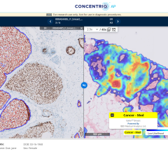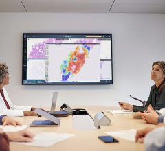
VividLook software helps spot cancer by imaging the speed of contrast flow into and out of the prostate.
Perhaps one of the most frightening experiences a patient can have is an inconclusive cancer test, where the biopsy is negative, but several other signs point to the possibility that the disease may be present. For some prostate cancer patients, this frightening scenario is a reality.
Patients who have a negative biopsy, but a high prostate-specific antigen (PSA), may be placed in a “watchful waiting” category, where they have to wait another six months between blood tests.
Jason Mullinix, M.D., director of body imaging at Indianapolis’ Franciscan St. Francis Health, said such an experience can take its toll on a patient.
“That’s pretty traumatic on an individual to go six months to a year between blood tests, all the while wondering, ‘do I have cancer or not?’” he said.
However, thanks to computer-aided detection (CAD) software, that has all changed.
CAD software helps doctors interpret medical images, especially when images from X-ray, magnetic resonance imaging (MRI) or ultrasound scans may be difficult to read. Typically, the program will highlight tumors or other suspicious areas.
Mullinix and his team at St. Francis – a multi-campus, 400-bed facility on the south side of Indianapolis – started using iCAD’s VividLook for prostate cancer in January 2010. Part of the reason for the switch to the CAD software was because St. Francis acquired new technology. Previously, the hospital used an endo-rectal probe. But with a new 32-channel magnet, the facility could perform prostate MRI imaging without it.
Uncovering Cancer
Although prostate MRI is nothing new, Mullinix said combining it with the CAD software drastically improved diagnostic quality.
“Prostate MRI has been around for awhile,” he said. “But the CAD takes it from around the 70-80 percent sensitivity/specificity into the 90s.”
This has been especially important for Mullinix and his team because prostate cancer can be particularly tricky to detect. To begin with, the organ itself can make imaging and detection difficult. On medical scans, the prostate appears solid – unlike the breast, which is mainly fat – making the detection of tumors or masses more challenging. Additionally, prostate cancer is slow-growing, which can sometimes make it hard to distinguish from adjacent healthy tissue.
As a result, Mullinix uses the CAD software primarily for “diagnostic augmentation.” When examining a potential prostate cancer patient, they will take three sequences, two of which do not use CAD. If the CAD image doesn’t line up with the other two, Mullinix knows further investigation is needed. If all three line up, then he knows, with a high degree of certainty, that therapy is needed.
How It Works
VividLook relies on the same principles with prostate cancer as it does with breast cancer. Like any other type of cancer, prostate cancer developes new blood vessels to deliver food and other nutrients. However, because the vessels are made quickly, they are often porous, and the software exploits this trait.
Contrast is injected into the patient, and clinicians monitor how fast it flows into and out of the prostate.
“If the patient has leaky blood vessels, then there’s a fast wash into the prostate tissue and a fast wash out,” Mullinix said. “And if it washes in quickly and leaves quickly, the computer program will color that red.”
Similarly, if the wash in is quick, but the wash out is slow, the program colors those areas blue or green. The program allows Mullinix to better target his biopsies. Typically, prostate cancer biopsies are taken from random areas of the prostate in order to get a good sample. However, the CAD software lets him take some of the guesswork out of the sampling.
Even though St. Francis has only been using the software since January, Mullinix says it has already played a vital role in helping several patients.
In one case, a patient came in with a high PSA, which is a common marker for prostate cancer. However, to complicate matters, the patient was also on blood thinners for a heart valve problem. As a result, the team wanted to avoid doing the standard random biopsies. Mullinix and his team decided to use MRI with the CAD software, with surprising results.
“We were able to localize a very small area that had a high wash in and a high wash out, and it was positive on the other two sequences as well,” he said.
Thanks to the software, they not only knew where to biopsy, they also knew the high PSA most likely wasn’t due to an infection and cancer was a distinct possibility. As a result, they decided it was best to briefly take the patient off his blood thinners to do the biopsies.
Even more recently, the software potentially helped change a patient’s therapy.
This patient had undergone chemo and radiation therapy to treat rectal cancer, and the follow-up MRI came out negative. Several doctors looked at the images and concluded they were negative. At the last minute, though, one of the clinicians recommended using VividLook. So the patient was injected with contrast.
“We imaged him again and sure enough, in the folds where it’s very hard to see, the cancer was hiding,” Mullinix said.
Peace of Mind
The CAD software is also useful in tracking the effectiveness of cancer treatment. As radiation or chemo is administered – and subsequently as those porous blood vessels are killed – the clinician can track the highlighted area as it turns from red to green and finally to blue.
Perhaps most important, though, is the effect that the software can have on a patient. It can’t quite exclude the presence of cancer yet. But the results, whether positive or negative, can help relieve a patient’s stress and anxiety during an extremely distressing time.
“It just gives you an extra sequence and extra chance of finding that cancer,” Mullinix said. “It just adds a level of peace of mind.”


 July 26, 2024


