In a rare showing of bipartisan support, on April 14, 2015, Congress repealed the sustainable growth rate (SGR) and approved a new framework to allow Medicare to transition from its current fee-for-service payment system to a value-based reimbursement system. The legislation (H.R. 2) eliminates the threat of annual cuts in Medicare payments to providers that were required under the SGR, which was approved more than a decade ago to control skyrocketing Medicare costs.
Established more than 30 years ago, Hawaii Radiologic Associates (HRA) has grown from its modest beginnings as Hilo Radiologic Associates to the Big Island of Hawaii’s most advanced radiology group. Today, the group of nine specialty trained radiologists provides diagnostic imaging services in four outpatient imaging centers located in East and West Hawaii and also provide professional interpretation services at four hospitals/medical centers around the island.
As the saying goes, sometimes less is more — a maxim that is proving true in the world of medical imaging as remote viewing systems continue to advance. While some manufacturers are still utilizing software-based systems for reading and sharing imaging data, many are embracing browser-based models, otherwise known as zero-footprint viewers.
Fujifilm’s APERTO Lucent is a 0.4T mid-field, open MRI system addressing today’s capability and image quality needs ...
Perhaps the biggest trend in health information technology today is the movement away from traditional picture archiving and communications systems (PACS) and cardiovascular information systems (CVIS) to enterprise imaging and data management systems. This migration is important, because it will fundamentally change how clinicians have been accessing images and patient information for the past 20 years.
Proton therapy has been on the rise in recent years, both in the United States and abroad, with more and more proton centers opening their doors to cancer patients around the globe. It provides an attractive alternative to conventional radiation therapy, as the directed beam more intensely attacks cancerous tissue while sparing surrounding healthy tissue and organs. In addition, radiation oncologists can control the depth to which the proton beam penetrates, and the beam will not travel any further, unlike traditional X-ray-based radiation therapy.

SPONSORED CONTENT — Fujifilm’s latest CT technology brings exceptional image quality to a compact and user- and patient ...
There have been several technology advances in PET/CT (positron emission tomography/computed tomography) systems recently introduced by vendors. On the show floors of the 2014 Society of Nuclear Medicine and Molecular Imaging (SNMMI) meeting and the 2014 Radiological Society of North America (RSNA) meeting, Philips, Toshiba and GE Healthcare introduced new PET/CT systems.
SPONSORED CONTENT — Fujifilm’s latest CT technology brings exceptional image quality to a compact and user- and patient ...
It’s the invisible elephant in radiology — radiation exposure. As physicians, we know all too well that exposure to radiation should be limited to the least amount possible. We have all learned about the ALARA principle for keeping exposure low. We also take precautions on our patients’ behalf and educate them. But what about us? The physicians and technical staff who treat patients every day cannot avoid radiation exposure entirely. However, there is growing evidence that not only is radiation exposure detrimental to our health, but also many long-term cumulative effects are still unknown.
The ITN team has just returned from the Healthcare Information and Management Society’s (HIMSS15) annual conference in Chicago, where more than 43,000 healthcare IT professionals gathered to learn about and share information on optimizing healthcare outcomes using information technology.
The ITN team has just returned from the Healthcare Information and Management Society’s (HIMSS15) annual conference in Chicago, where more than 43,000 healthcare IT professionals gathered to learn about and share information on optimizing healthcare outcomes using information technology.
SPONSORED CONTENT — EnsightTM 2.0 is the newest version of Enlitic’s data standardization software framework. Ensight is ...
Based on its ongoing analysis of the medical imaging market, Frost & Sullivan recognizes Toshiba America Medical Systems, Inc. with the 2015 North American Frost & Sullivan Award for Medical Imaging Company of the Year. The company has changed the way it approaches the imaging market by offering scalable, upgradable technologies and a patient-focused view of partnerships. This value-based approach positions the company for continued success in the medical imaging market by allowing it to re-orient many of its imaging customers away from a cyclical sales model and toward deeper, more renewable strategic partnerships.
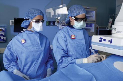
Texas Health Presbyterian Hospital Plano is the first hospital in North Texas to offer full-body three-dimensional imaging with a lower dose of radiation than traditional imaging.
One life-threatening complication of lung cancer surgery is the formation of blood clots in the lungs (also called pulmonary embolism, PE) or in the legs (also known as deep vein thrombosis, DVT). Together, they would be defined as venous thromboembolic events (VTE). Several presentations at AATS 2015 shed new light on this serious problem. In the first prospective study of its kind, the incidence of VTE was found to be higher than previously reported, with a 5.4 percent VTE-specific mortality rate. Of concern to clinicians, most events were asymptomatic and occurred after patients were discharged from the hospital. The second report highlights the importance of screening for VTEs, especially since the majority of lower extremity VTEs found after pneumonectomy would have gone undiagnosed and untreated without screening. The third report describes a risk assessment tool for VTEs that is applied for the first time to predict an individual’s risk of VTEs after lung cancer surgery, which can help clinicians decide whether prolonged anti-clotting therapy is warranted.
Did you know that approximately one-third of all the data in world is created by the healthcare industry and that ...
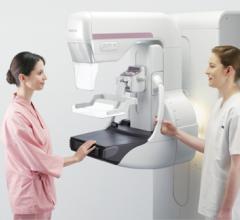
The U.S. Food and Drug Administration (FDA) is alerting patients who had mammograms at Coastal Diagnostic Center in Pismo Beach, California, on or after Feb. 24, 2013, about possible problems with the quality of their mammograms. This does not mean that the results of the mammograms were inaccurate, but it does mean that the patients should consider having their mammograms re-evaluated at a Mammography Quality Standards Act (MQSA)-certified facility to determine if they need a repeat mammogram or additional medical follow-up.
Proton Partners International Limited announced the appointment of global partners in its plans to build three proton beam therapy centers in the U.K. Partner organizations will provide state-of-the-art clinical equipment and technology solutions to the company.
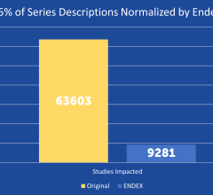
SPONSORED CONTENT — EnsightTM 2.0 is the newest version of Enlitic’s data standardization software framework. Ensight is ...
Two new studies show that increasing the dose of radiotherapy given to children with an intracranial ependymoma, a form of cancer of the central nervous system, can significantly improve their survival. Results of both studies were presented at the third European Society for Radiotherapy and Oncology (ESTRO) Forum in Barcelona, Spain.
The American College of Radiology (ACR) announced its support for the Diagnostic Imaging Services Access Protection Act (H.R. 2043) which seeks to prospectively repeal the existing 25 percent Multiple Procedure Payment Reduction (MPPR) applied to Medicare reimbursement. The MPPR is applied for interpretation of advanced diagnostic imaging scans performed on the same patient, in the same session, on the same day.
A first-in-human prostate cancer study in the Journal of Molecular Imaging and Biology showed initial safety, biodistribution and dosimetry results with [18F]DCFPyL, a second-generation fluorine-18 labeled small-molecule prostate-specific membrane antigen (PSMA) inhibitor. The imaging biomarker has been developed at Johns Hopkins University in Baltimore by study co-author Martin G. Pomper, M.D., Ph.D.
Ultrasound has become an essential tool for diagnostic and procedural uses in the critical care environment. The May issue of Anesthesia & Analgesia outlines a new set of basic ultrasound learning skills developed specifically for use in anesthesiology-critical care medicine (ACCM) training programs.
Kaiba was just a newborn when he turned blue because his little lungs weren’t getting the oxygen they needed. Garrett spent the first year of his life in hospital beds tethered to a ventilator, being fed through his veins because his body was too sick to absorb food. Baby Ian’s heart stopped before he was even six months old.
With higher deductibles and more price-conscious healthcare consumers, as a result of the Affordable Care Act, improving the performance, comfort and speed of MRI scans can be an important strategy for imaging centers that must compete by offering the best patient care and experience.


 May 04, 2015







![prostate cancer, [18F]DCFPyL, PSMA, biomarker, Johns Hopkins, WMIS](/sites/default/files/styles/content_feed_medium/public/Prostate%20cancer%20F18.jpg?itok=QYOFHfKK)


