Case Study | Agfa HealthCare
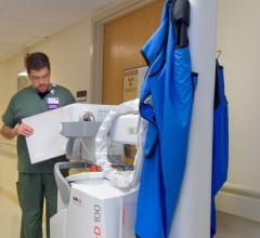
Upgrading its technology was a big decision for US Signature HealthCare in Brockton, Mass. The oldest and largest in-patient hospital within its area, its ambitious upgrade program covered a lot of goals; including offering digital imaging in the emergency room and providing more tools to the people making time-sensitive clinical decisions. In rigorous field trials, one solution stood out, offering not only the specifications the hospital wanted but also visibly different image quality: the Agfa HealthCare DX-D 100. And that was just the start!
 November 05, 2013
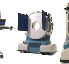
News
With a strong foundation of expertise in imaging design, development and manufacturing, NeuroLogica transforms fixed technologies into portable point-of-care platforms. The current product offering consists of three portable platforms, the CereTom small bore CT, inSPira HD brain SPECT and the BodyTom large bore CT.
November 05, 2013 
News
Viztek announced a new partnership with Matakina to integrate the company’s VolparaDensity software for analyzing digital mammography and tomosynthesis images and objectively assessing breast density with the Viztek Opal-PACS and Opal-wRIS for streamlined reporting and workflow.
November 04, 2013 Sponsored Content
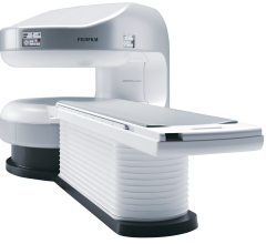
Fujifilm’s APERTO Lucent is a 0.4T mid-field, open MRI system addressing today’s capability and image quality needs ...
September 25, 2024 
Blog
Annoyed by the high cost of a tune-up, 30 years ago I took a class on automotive maintenance. Armed with a spark plug wrench, timing light and screwdriver, I thereafter tuned up — far more often than necessary — my 1978 Toyota Corolla. Back then I had the right tools and the knowledge to do what needed to be done. Today I have neither. Radiologists are in a different ocean, but the same boat.
November 04, 2013 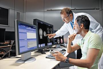
Feature | Greg Freiherr
Annoyed by the high cost of a tune-up, 30 years ago I took a class on automotive maintenance. Armed with a spark plug wrench, timing light and screwdriver, I thereafter tuned up — far more often than necessary — my 1978 Toyota Corolla. Back then I had the right tools and the knowledge to do what needed to be done. Today I have neither.
November 04, 2013 Sponsored Content

News | Computed Tomography (CT) | August 06, 2024
SPONSORED CONTENT — Fujifilm’s latest CT technology brings exceptional image quality to a compact and user- and patient ...
Feature | Anders Granlund
Digital breast tomosynthesis (DBT) was approved in the United States for use as a supplement to traditional mammography following U.S. Food and Drug Administration (FDA) review of two studies in which radiologists showed a 7 percent improvement in the ability to distinguish between cancerous and noncancerous cases using 3-D datasets.
November 04, 2013 Sponsored Content
News | Computed Tomography (CT)
SPONSORED CONTENT — Fujifilm’s latest CT technology brings exceptional image quality to a compact and user- and patient ...
August 06, 2024 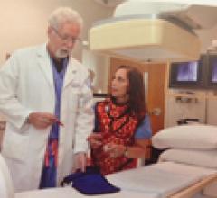
Case Study | Sponsored by GE Healthcare
The Hospital for Special Surgery (HSS) in New York, N.Y., consistently ranks No. 1 on the U.S. News & World Report list of best orthopedic hospitals in the United States. The hospital performs thousands of surgeries every year, and those procedures require leading-edge imaging technology. HSS currently uses 20 GE OEC fluoroscopy systems in its operating suites. That’s a lot of imaging, and a big challenge for a department whose motto is “every procedure starts with low dose.”
November 04, 2013 
Feature | Dave Fornell
As the use of mobile computing devices and smartphones has rapidly proliferated in healthcare over the past few years, there has been a flood of medical applications (apps) developed for all facets of medicine. In an increasingly tech savvy world, people want information to be at their fingertips when and where they need it via their mobile devices.
November 04, 2013 
Feature | Williette Nyanue
The Robert H. Lurie Comprehensive Cancer Center of Northwestern University is one of 41 National Cancer Institute (NCI)-designated “comprehensive” cancer centers in the country — the highest ranking given by the NCI. The Lurie Cancer Center was first established at Northwestern University in 1974, and is dedicated to scientific discovery, providing state-of-the-art care, and training clinicians and scientists.
November 04, 2013 Sponsored Content
News | Artificial Intelligence
SPONSORED CONTENT — EnsightTM 2.0 is the newest version of Enlitic’s data standardization software framework. Ensight is ...
June 21, 2024 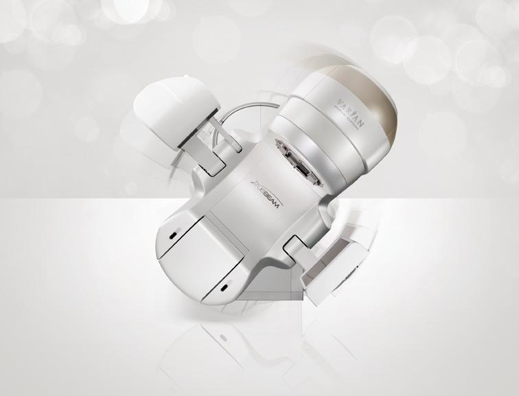
Feature | Williette Nyanue
Radiation therapy has played an important role in the treatment of cancer for more than a century. Used typically as a curative treatment either alone or in conjunction with surgery and/or chemotherapy, the aim of radiation therapy has always been to eradicate a patient’s cancer.
November 04, 2013 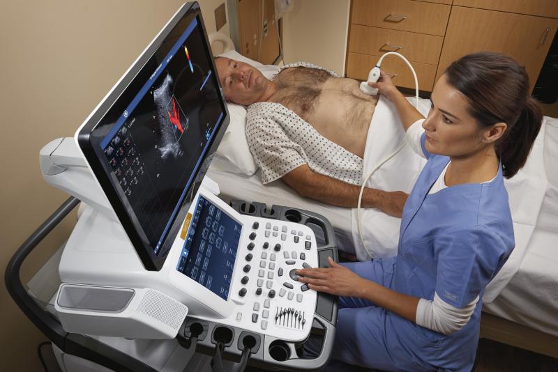
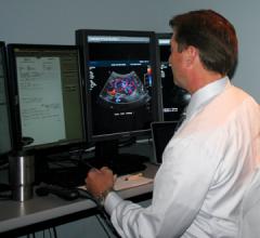
Feature | Ultrasound Imaging | Williette Nyanue
Physicians have been utilizing conventional ultrasound, also known as b-mode ultrasound, for diagnostic imaging since the 1970s. However, over the past 10 years there have been significant technological improvements within the equipment, as well as development of new technologies that allowed ultrasound to become more widely adopted. Ultrasound equipment has gotten physically smaller, generates less heat and has become more power efficient. These upgrades, along with vast enhancements in image quality, have pushed ultrasound into the point-of-care setting. Point-of-care ultrasound has become widely performed in emergency rooms, PCP offices and obstetric practices. As healthcare reform continues to favor the use of more cost-effective solutions, this trend is expected to persist until ultrasound is used in every doctor’s office.
November 04, 2013 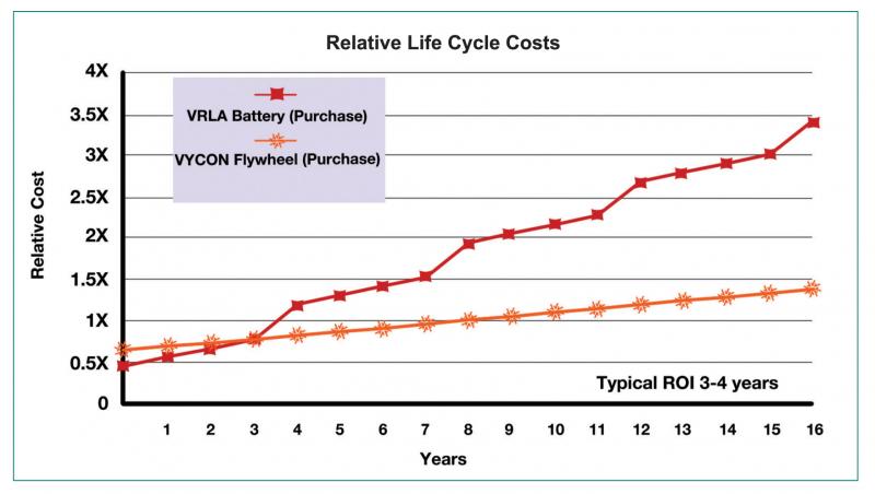
Feature | Frank DeLattre
Whether it is a magnetic resonance imaging (MRI), computed tomography (CT) or another type of imaging system, reliable power protection is critical for the proper operation and uptime of imaging applications. But what happens if a healthcare facility suffers a power brownout, surge or outage? How do these events affect imaging equipment?
November 04, 2013 Sponsored Content

Feature | Artificial Intelligence
Did you know that approximately one-third of all the data in world is created by the healthcare industry and that ...
June 03, 2024 
Case Study | Sponsored by EIZO Inc.
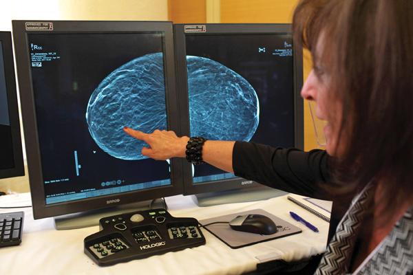
Elizabeth Wende Breast Care in Rochester, N.Y., is internationally recognized as a leader in the field of breast imaging and breast cancer diagnosis. It is one of the largest freestanding breast imaging centers in the United States with American College of Radiology (ACR) accreditation and U.S. Food and Drug Administration (FDA) certification.
November 04, 2013 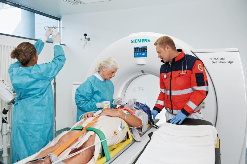
Feature | Computed Tomography (CT) | Raissa Rocha
With concerns about radiation dose and reducing unnecessary imaging scans, advances in computed tomography (CT) systems have brought about technologies such as iterative reconstruction software, intraoperative capabilities and dose-tracking software. In addition, recent studies on the use of CT on select patient populations and the modality’s benefits in detecting certain cancers are showing that the risks of CT imaging can go both ways. While CT exams can add to a patient’s lifetime exposure to ionizing radiation, they can also be more beneficial in cases where magnetic resonance imaging (MRI) or ultrasound might not be able to detect early-stage cancers. Some of these trends in utilization indicate that appropriate low-dose CT imaging will be key across patient populations.
November 04, 2013 Sponsored Content
News | Artificial Intelligence | June 21, 2024
SPONSORED CONTENT — EnsightTM 2.0 is the newest version of Enlitic’s data standardization software framework. Ensight is ...
Case Study | Sponsored by PACSGEAR
Voice recognition and template-driven radiologist reporting have dramatically streamlined delivery of the clinical report. However, including measurement data in a report still requires radiologist dictation of numbers manually captured by a technologist. Since quantitative measurements are an important and common aspect of the radiologist report, further increases in productivity and clinical quality can be realized by automating this process.
November 04, 2013 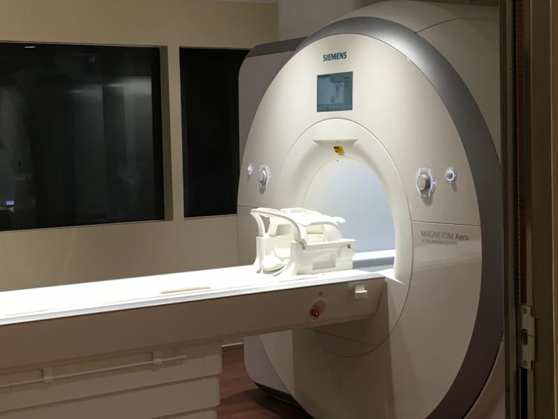
Feature | Magnetic Resonance Imaging (MRI) | Williette Nyanue
Wide bore magnetic resonance imaging (MRI) systems have allowed radiologists to offer patients the optimized comfort of conventional open bore systems, as well as the high-quality imaging of conventional closed bore systems. Because wide bore MRIs have broadened the demographic of patients who can be tested, the systems have gained widespread adoption in use, with many practices opting to equip their offices solely with wide bore systems.
November 04, 2013 
Case Study | Sponsored by Hologic, Inc.
By all measures the Northwest Indiana Breast Care Center at Methodist Hospitals is a success. Opened in 2012, the Breast Care Center embodies Methodist Hospitals’ commitment to provide the most advanced technologies available for the early detection and treatment of breast cancer. Methodist Hospitals is a not-for-profit, community-based healthcare system with two full-service campuses located 14 miles apart in Gary and Merrillville, Ind.
November 04, 2013 
Feature
Clinical trial results demonstrated that a noninvasive coronary computed tomography angiography (CTA)-based test accurately assesses coronary artery disease (CAD) with results closely matching those of invasively measured fractional flow reserve (FFR), and may inform potential revascularization treatment options, including angioplasty and coronary artery bypass surgery (CABG), better than current methods.
November 01, 2013 News
Image IQ, Inc. announced that findings from an orthopedic clinical study conducted by ImageIQ and Cleveland Clinic show that professional baseball pitchers with lower degrees of dominant humeral torsion, or the degree of twisting of the long arm bone running from shoulder to elbow, are more prone to severe arm and shoulder injuries.
November 01, 2013 Blog
By Melinda Taschetta-Millane, editorial director
October 31, 2013 © Copyright Wainscot Media. All Rights Reserved.
Subscribe Now