News
October 7, 2011 — U.S. Food and Drug Administration (FDA) Commissioner Margaret A. Hamburg, M.D., released a blueprint containing immediate steps to drive biomedical innovation, while improving the health of Americans.
 October 07, 2011
 Blog
When it comes to medical imaging, pick any part of the body other than the female breast and the FDA pays little notice. This particular part of the anatomy gets an extraordinary amount of attention, particularly as it pertains to cancer. Politics has a lot to do with it.
October 06, 2011 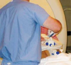
Case Study
Radiologist John Feller, M.D., medical director of Desert Medical Imaging in Indian Wells, Calif., and local urologists have joined forces to test a promising new way to detect and diagnose prostate disease. When a traditional transrectal ultrasound (TRUS) biopsy proves negative for the presence of disease, yet a patient’ s prostate-specific antigen (PSA) levels continue to rise, magnetic resonance imaging’s (MRI) excellent soft-tissue imaging quality may be the answer to a difficult diagnosis.
October 06, 2011 Sponsored Content
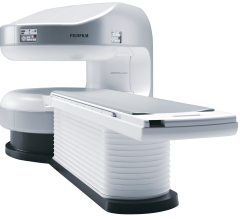
Fujifilm’s APERTO Lucent is a 0.4T mid-field, open MRI system addressing today’s capability and image quality needs ...
September 25, 2024 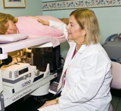
Case Study
The sign outside the Women’s Center for Radiology in Orlando, Florida, announces the arrival of 3D mammography. Susan Curry, M.D., founder and medical director, wants to get the message out to women in Central Florida about 3D mammography and the difference it can make in the early detection of breast cancer.
October 06, 2011 
Feature | Howard Reis
While determining how to best measure quality in a teleradiology operation is more of an art, rather than a science, I want to propose the following equation: TQ = fn (CV + QA% + TAT + QoS). In this equation, teleradiology quality is a function of the credentials of the reader (CV), the miss rate (QA%), the turnaround time for studies (TAT) and the overall quality of service (QoS) delivered.
October 06, 2011 Sponsored Content

News | Computed Tomography (CT) | August 06, 2024
SPONSORED CONTENT — Fujifilm’s latest CT technology brings exceptional image quality to a compact and user- and patient ...

Case Study
With the need to transport images and make imaging studies readily available to referring physicians, WCGH got into the picture archiving and communications system (PACS) game relatively early, implementing its first system in 2003. When it contacted Infinitt North America (then SmartPACS) at that time, no one knew it would be the start of a technology partnership that would support them into the next decade or that its PACS would become the hub of its clinical IT platform.
October 06, 2011 Sponsored Content
News | Computed Tomography (CT)
SPONSORED CONTENT — Fujifilm’s latest CT technology brings exceptional image quality to a compact and user- and patient ...
August 06, 2024 
Case Study
“The time we save is priceless,” said Thomas G. Frazier, M.D., medical director of the Comprehensive Breast Center at the Bryn Mawr Hospital, referring to their use of the KUBTEC XPERT 40 specimen radiography system in the operating room. A nationally recognized surgical oncologist specializing in breast cancer, he performed the first immediate breast reconstruction surgery in the Philadelphia area. He uses the XPERT 40 at Bryn Mawr Hospital, “at least four or five times a week, maybe more, in the operating room alone!”
October 06, 2011 
Feature | Helen Kuhl
Although computerized physician order entry (CPOE) systems have been around for a few years, only a few vendors really embraced the market with robust offerings and only a few healthcare providers showed interest in adopting them — until the 2009 passage of the American Reinvestment and Recovery Act (ARRA). After that, the landscape changed considerably, as many providers wanted to take advantage of reimbursements available in meeting meaningful use (MU) requirements and the rate of adoption grew significantly.
October 06, 2011 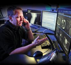
Case Study
Desert Radiologists is a high-efficiency practice in Las Vegas, Nev., that performs 1.25 million exams per year with a staff of 45 radiologists. Yet the practice operated with an outdated and overloaded picture archiving and communications system (PACS) that had no common worklist, no flexibility and could no longer handle its daily imaging volume. Additionally, as Desert Radiologists’ imaging volume continued to increase, its existing storage solution could not keep up. That system was a vendor-neutral archive with on-site SAN/NAS units. However, sending data to the vendor-neutral archive created two PACS and increased the complexity of managing and purging the data.
October 06, 2011 Sponsored Content
News | Artificial Intelligence
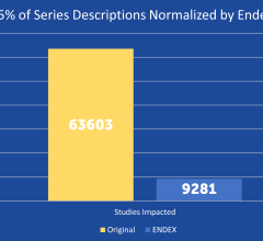
SPONSORED CONTENT — EnsightTM 2.0 is the newest version of Enlitic’s data standardization software framework. Ensight is ...
June 21, 2024 
Feature | Michael S. Gossman, M.S., DABR and David Coll-Segarra, M.S.
New research shows the effects of electron beams on implanted vascular access ports composed of plastic, determining how they impair the fluence of radiation around them.
October 06, 2011 
Feature | Vivek Mehta, M.D.
One of the most important recent advances in radiation oncology has been the integration of 4-D treatment planning tools into the clinic. 4-D treatment tools have enabled the radiation oncologist to better plan and account for the tumor motion in a specific patient.
October 06, 2011 Feature | Tobias Gilk
Magnetic resonance imaging (MRI) is safe, profoundly safe, provided we follow industry best practices. Unfortunately, the rates of reported MRI accidents are nearly five times what they were just five years ago, according to data from the U.S. Food and Drug Administration (FDA)[1] — an apparent indication that we, as an industry, aren’t following best practices.
October 06, 2011 Sponsored Content

Feature | Artificial Intelligence
Did you know that approximately one-third of all the data in world is created by the healthcare industry and that ...
June 03, 2024 
Feature | Helen Kuhl
With healthcare professionals thinking about the effects of ionizing radiation on the population at large, there is particular concern about its use for imaging children. That there is reason for concern was underscored by the release of new study results earlier this year, which indicated computed tomography (CT) exams of children in hospital emergency departments increased substantially from 1995 to 2008. While CT still may be the best imaging choice in certain cases, there is continued emphasis on using other modalities whenever possible. As a result, magnetic resonance imaging (MRI) is being used more and more,
October 06, 2011 Feature | Roberto G. Aranibar, Frost & Sullivan Healthcare Research Analyst
The overarching trend in ultrasound continues to be the development of smaller and more powerful imaging platforms. The provision of hand-carried systems that offer advanced functionality and premium image quality in a small, easy-to-use and affordable package has almost become essential to growth in ultrasound over the last few years.
October 06, 2011 Sponsored Content
News | Artificial Intelligence | June 21, 2024
SPONSORED CONTENT — EnsightTM 2.0 is the newest version of Enlitic’s data standardization software framework. Ensight is ...
Feature | Mary Beth Nevulis
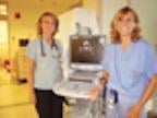
The number of ultrasound systems in emergency departments will nearly double by 2015, according to a report by ultrasound industry expert and consultant Harvey Klein. One medical professional who has had success using ultrasound in the emergency department is Colleen Campbell, M.D., a professor of emergency medicine and the director of emergency ultrasound at the University of California San Diego.
October 06, 2011 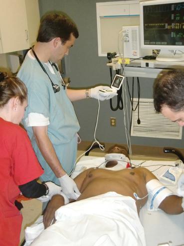
Feature | Thomas Cook, M.D.
Thomas Cook, M.D., is the emergency medicine residency program director at Palmetto Health Richland in Columbia, S.C., and has been using ultrasound in the practice of emergency medicine since 1996. He also had the recent experience of traveling in China and learning about that country’s use of ultrasound. He shares his expertise and views about ultrasound in the emergency department (ED) in the following Q&A with ITN.
October 06, 2011 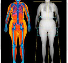
Technology
October 5, 2011 — Hologic will display the new Advanced Body Composition assessment feature of its Discovery Dual Energy X-ray Absorptiometry (DXA) system at RSNA 2011.
October 05, 2011 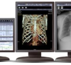
Technology
During RSNA 2011, Infinitt will showcase its latest advances in image and information management across the enterprise.
October 05, 2011 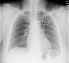
Technology
Riverain’s OnGuard chest X-ray computer-aided detection (CAD) software helps identify nodules that may be early-stage ...
October 05, 2011 Technology
October 5, 2011 — Fukuda Denshi USA has announced the introduction of the company's new UF-760AG ultrasound system in the United States. The color portable unit provides imaging quality in a compact system and is designed for diagnostic use in numerous specialty markets.
October 05, 2011 © Copyright Wainscot Media. All Rights Reserved.
Subscribe Now