May 18, 2023 — Royal Philips, a global leader in health technology, announced the launch of the Philips CT 3500, a new high-throughput CT system targeting the needs of routine radiology and high-volume screening programs. Powered by AI, the Philips CT 3500 includes a range of image-reconstruction and workflow-enhancing features that help to deliver the consistency, speed, and first-time-right image quality needed for confident diagnoses by clinicians and increased return on investment – even in the most demanding, high-volume care settings.
“Increased financial pressures, chronic staff shortages, and escalating patient demand are driving radiology departments to do everything they can to maximize throughput, to guarantee equipment uptime, and to avoid repeat scans,” said Frans Venker, General Manager Computed Tomography at Philips. “Today, many radiology departments scan hundreds of patients a day. We’ve engineered the Philips CT 3500 to reduce the pain points that these high-volume departments face by developing a versatile, reliable, high-throughput imaging solution. It automates radiographers’ most time-consuming steps so that they can spend more time focusing on the patient.”
Accelerating workflows
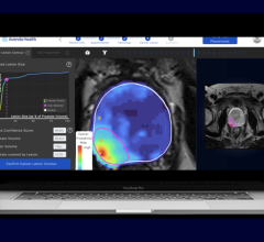
The CT 3500 features Philips’ latest AI-powered CT Smart Workflow to automate every step in the scanning process. Precise Position uses a camera to automatically determine patient orientation, improving positioning accuracy by 50% while reducing patient positioning time by up to 23% [1]. Precise Planning automatically determines the area to be scanned and the appropriate Exam Card based on the patient’s anatomy. This provides fast exam preparation and can improve inter-operator consistency. Precise Intervention can offer automated setup and treatment guidance for tissue biopsies and other needle-based interventions.
Delivering high-quality images at low dose
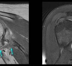
Precise Image AI-based reconstruction is designed to deliver the high image quality needed by radiologists to make a precise diagnosis. Precise Image allows radiology departments to simultaneously achieve up to 60% improved low-contrast detectability, 85% lower noise, and 80% lower radiation dose. All reference protocols are reconstructed in under a minute to support even the busiest radiology departments [2,3].
Boosting up-time
The CT 3500 is designed to deliver the uninterrupted imaging required by high-throughput radiology departments and screening programs including mobile screening units. To achieve this level of reliability, the CT 3500 is built on Philips’ highly regarded vMRC tube and tracks critical performance metrics with internal and external proactive monitoring sensors that allow Philips service engineers to intervene prior to any potential impact on CT operations.
Adding to Philips’ existing portfolio of high-performance CT solutions, the CT 3500 is being launched back-to-back at the 2023 China International Medical Equipment Fair (CMEF 2023, May 14 - 17, Shanghai, China) and the 2023 Deutscher Röntgenkongress (ROKO 2023, May 17 -19, Wiesbaden, Germany).
For more information: www.philips.com
References:
[1] Based on Philips in-house assessment by five clinical experts, comparing manual versus Precise Positioning in 40 clinical cases using a human body phantom. Results from case studies are not predictive of results in other cases. Results in other cases may vary.
[2] In clinical practice, the use of Precise Image may reduce CT patient dose depending on the clinical task, patient size, and anatomical location. A consultation with a radiologist and a physicist should be made to determine the appropriate dose to obtain diagnostic image quality for the particular clinical task. Dose reduction assessments were performed using reference body protocols with 1.0 mm slices at the “Smoother” setting of Precise Image, and tested on the MITA CT IQ Phantom (CCT189, The Phantom Laboratory) assessing the 10 mm pin and compared to filtered-back projection. A range is seen across the 4 pins, using a channelized hoteling observer tool, that includes lower image noise by 85% and improved low-contrast detectability from 0% to 60% at 50% to 80% dose reduction. NPS curve shift is used to evaluate image appearance, as measured on a 20 cm water phantom in the center 50 mm x 50 mm region of interest, with an average shift of 6% or less.
[3] Precise Image is currently not available for pediatrics.
 May 20, 2024