Decades since the advent of breast scanning technology, innovations in noninvasive diagnostic imaging provide new options in the field of early detection. A mammogram can show how dense breasts are including how low or high in density. However, overcompression artificially lowers the radiographic density.

Image 1.
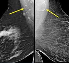
In this standard mammogram (see Image 1), a dense breast is presented side by side. The white shaped “V” that comes down the top center are the pectoral muscles of the chest wall. On the far outside, the white line is the skin outline of the breast. This is the dermal tissue causing the white line viewed enface. Radiologists always study this for any indication of inflammatory disease of the skin or inflammatory breast cancer, which manifests itself in skin thickening. Between the center wedge and the skin outline, you will find homogeneous cloudy areas with patchy black spaces within as an example of dense breast tissue.

Image 2.
Usually, dense breast tissue appears white on a mammogram. We must identify them as one of two forms of breast density; one is called fibrocystic or fibrous (see Image 2) which is homogeneously white. Occasionally you can see a branching of blood vessels, dilated ducts or a streak of fat inside the dense breast tissue. This is the most common type of dense breast tissue and generally seen in the over 40 population.

Image 3.
Another example of a dense breast shows the difference between homogeneous white versus the whitish area (see Image 3). This is filled with dark, wormy looking structures, which are the breast glands called glandular tissue. This kind secretes milk and its glands are often dilated. Both fibrous and glandular may appear similar under a mammogram as highly dense areas, but they look completely different under an ultrasound scan. Through ultrasound, we can check for tumors easily through fibrotic dense breasts because it stands out as a black region (or a black hole) within the white area. As shown in Image 3, a black hole could get lost, making it more difficult to image this type of dense breast. In this case, a solution is the use of elastography (see Image 4), which offers visual confirmation as indicated by color data. Elastography can measure tissue density (its hardness or elasticity) within the glandular breast tissue.

BRCA Positive in Glandular Density.
This tissue type is more common in the under-40 age group and is associated with other glandular proliferation such as endometriosis and is reportedly linked to dermal inflammation. In published reports, comparative studies between fibrous and glandular breast tissue studies remain limited. We are observing (especially in the younger age groups) expanding reviews of these types of tissue density aligned with the rates of breast cancer to confirm the rate of malignancy in tissue alteration from normal.

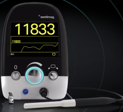
Image 4: Elastography of breast mass.
Remote Imaging Collaboration of Ultrasound Breast Scanning
Since the COVID-19 pandemic, I designed and established a remote imaging paradigm, called TeleMed Scans, with researchers and clinicians abroad and in other states. I found the current digital imaging solutions to be quite Wi-Fi friendly and ready for virtual collaboration. This widely expanded my capacity to further support ultrasound imaging in the global front lines.

I took on the role of diagnostic imaging investigator for a dense breast screening program that conducted scans from various remote locations. I received all scans on my desktop and generated analytical reports to identify or confirm breast density and breast health of all the test subjects acquired. Our partnership showed the flexibility of 3D ultrasound for screening of breast tissue condition and potential disease. The screening sites targeted women under 39, and women with dense breasts, which was theorized to be more common in underserved areas and younger women (below the age of standardized screening).
Portable Scanners
Upon review of a wide range of portable ultrasound devices, one model came to mind; my field partners often hear me discuss the Terason T-3200 due to its proven quality, user-friendly interface and its rugged design. Its capacity offers high resolution imaging and high quality probes that can capture anatomical scans comparable to hospital grade appliances. Hence, we are able to see not only the breast tissue, but also the skin. This means that if there’s a breast cancer that’s infiltrating the skin from below (which a clinician may not be able to see and oftentimes cannot feel) this will show that the cancer is a stage two because it’s already broken through the bottom layer of the skin. The Wi-Fi friendly model would capture images in real-time with quantitative accurate in finding cancers or benign cysts in dense or lumpy breast tissue.
As portable (field-based) ultrasounds go, including the hand-held models, device engineers have clearly captured the many needs of remote imaging equipment. From emergency response calls to triage situations, technicians have all expressed significant benefit in its intuitive user interface and the AI driven factory presents to streamline the screening process. Another one of my remote partners conducted breast cancer screening in a tent during a health event and reported the surprising hospital-grade quality and accuracy of today’s portables. The design of portable ultrasound units also respond to facilitating remote training.
Wi-Fi Supports the Medical Collaboration Paradigm
Today’s entire emergency response system is well underway the use of Wi-Fi connected devices in their emergency vehicles. The portable rescue services are completely reliant on Wi-Fi connections, allowing for field responders to receive medical guidance and diagnostic reading support from ‘central command’. Having access of the supervision of a senior medical professional on the field can save lives where precious seconds count — way before the patient arrives in the emergency room. The ability to have Wi-Fi allows the responder and the surgical team to confirm the pathology while receiving expert guidance from anywhere in the world within seconds.
An Observational Tour of Breast Density
Although breast density is a well-established and prevalent breast cancer risk factor, it’s biological connection is not (yet) clearly understood. More research is needed to support the population associated risk proportion in athletic pre-menopausal women. This quantitative data gathered from studies of women undergoing imaging ultrasound scans of pre-menopausal women with dense breasts.
Case 1: Normal Breast Tissue Image

This scan shows that directly under the white line (A) is a black area that is the epidermis of the skin. The horizontal white band below is the dermis. The resolution of the 3D ultrasound device allows us to see submillimeter lesions. Below this is an area (B) that’s mixed white and dark with horizontal bands of white and broad swaths of dark fatty tissue, white is breast tissue. The scan shows normal breast tissue as gray.
Case 2: Ultrasound Depth Capacity

To contrast from patient 1, we have a band of white tissue (C), in an area that normally has fatty breast tissue. Below C is the semi-circular bands represent the rib cage and more importantly, the surface of the lung. This is useful because should a cancer of the breast metastasize to the lymph nodes or to the lungs, this is where we might see evidence of this during this simple screening examination. The dense breast tissue is the band of white above the semi-circular rib cage echoes-in the middle of the band of whites, are tiny black wormy lines, which represent dilated ducts, which are a part of breast disease or cystic breast disease.
Case 3: Category 4 Density

The skin outline (epiderma layer) is on the top, with the curvilinear ribs in the middle of the scan as a broad white arc. Within the middle of this white path are tiny, bright white echoes representing micro calcium inside the breast. The fibrous tissue is an inflammatory process which heals with scarring and calcification that can also block and dilate the ducts. This an example of a very white dense breast with areas of micro calcification as an expected part of the healing process. This would be Category 4 breast density on mammography because the tissue is homogeneously white.
Case 4: Benign Tumor

Right below the word and the arrow is a black area within the white carpet of tissue that is sharply demarcated. It has echoes deep to this indicating high through transmission of sound energy as a white trail behind the black area showing that it’s a degenerated cystic area or a solid benign tumor that’s breaking down into cystic regions. This probably benign (BI-RADS 3) lesion is usually followed every six months with ultrasound scans. This would show up as a BI-RADS or as a class four density and probably be missed on mammograms. In a prior research project, we did a study showing that the benign tumors like this in dense breasts are missed about 90% of the time by mammography and they’re often hard to palpate clinically.
Case 5: Category 1 Density

This is an example of a one density mammogram or breast tissue, because this is almost uniformly dark or fatty tissue within the breast. The linear white lines are prominent (suporting fibrous septae) because the bulk of the tissue is predominantly fatty.
Case 6: Category 3 with Microcalcification

This is an example of a class three demonstrating white tissue interrupted by horizontal areas of dark and white indicating dilated ducts, some of which have micro calcification within them. Note the chest wall below the breast tissue so an infiltrating cancer could be imaged invading not only the skin, but the muscle tissue below that. With this technology accurate cancers staging is possible within minutes.
Case 7: Pectoral Muscle Imaging

This is another example of mixed density tissue with alternating bands of gray and darker gray. Below that is a very thick pectoral muscle below appearing as a white band dorsal to the rib cage. Muscle disease as well as the pleural disease (on the parietal lining of the lung). Measuring the thickness of a muscle is useful in treating muscle wasting diseases. During the COVID era occasionally pleural thickening and nodules were identified.
Early Detection is Key
Breast density is a key factor in early detection because mammograms routinely miss breast cancer in dense breasts, especially in younger and more athletic women. Ultrasound offers a supplemental scan and peace of mind because it finds the pathology and almost instantaneously distinguishes a benign cyst from a possible cancer — or a definite cancer. Ultrasound even goes further because if there is a definite cancer or something highly suspicious of a cancer image then staging may be initiated immediately at the other organs such as the glands under the arm to see if it’s spread into the lining of the lungs or invading the pleura or the lymph nodes in the abdomen or the brain. Since we are now armed with expert diagnostic over-readers and remote collaborative imaging options, I urge our gynecologic and obstetric community leaders to take advantage of this affordable non-invasive screening solution. One of the sessions at the global UltraCon 2024 symposium discussed the resistance to making screening ultrasound the norm for women under 40 and women with dense tissue. It is thought-provoking that in 2023 the nation of China switched from using mammography as the screening modality to high resolution ultrasound. Indeed, colleges and institutions of learning in the Northeast US are adopting teaching programs to educate the age group between 20-39 as well as underserved communities in the health advantages of sonographic screening. Some learning institutes are creating programs with AI to train class members to perform limited exams with video documentation and algorithms for further action.

Robert L. Bard, MD, PC, DABR, FASLMS, is an active medical director of Cancer Diagnostic Imaging Center (NYC), using advanced 3-D ultrasonographic Doppler imaging to detect cancers in breast, prostate, skin, thyroid, melanoma and other areas. He has extensive credentials as a clinical researcher/validator and has published medical textbooks and science journals. He is also a member of the ITN Editorial Advisory Board.
Author’s note: Special thanks to Terason Ultrasound and Mindray NA for their generous use of ultrasound imaging solutions included in this report. Additional thanks to Noelle Cutter, MD, and Alexandra Fiederlein, (Molloy University) and Abdellatif Zerzif (ultrasound and laser tech) for his stellar professional performance in managing both technologies and patient care. Also, extended thanks belong to research advisors Lennard Gettz, Roberta Kline, MD, and Leslie Valle-Montoya, MD, for their support throughout the coordination and execution of this review.
Related content:
ACR Issues Statement on Final USPSTF Breast Cancer Screening Recommendations
Task Force Issues Final Recommendation Statement on Screening for Breast Cancer
VIDEO: FDA Update on the US National Density Reporting Standard - A Discussion on the Final Rule
VIDEO: Research and Advancements in Breast Imaging Technology
One on One … with Wendie Berg, MD, PhD, FACR, FSBI
VIDEO: The Impact of Breast Density Technology and Legislation
 May 10, 2024