
Lorraine Drapek, DNP, nurse practitioner, radiation oncology, GI service, Massachusetts General Hospital, explains the roles of advanced practice providers in radiation therapy. She spoke on this topic at ASTRO 2019 at a session that reviewed the integration of APPs into radiation oncology practice to enhance clinical care. This includes but is not limited to: on-treatment management, symptom and acute toxicity management during treatment, inpatient consultations, procedural assistance, treatment planning, follow-up, survivorship and research.
itnTV
ON DEMAND WEBINAR: Strategies to Optimize Imaging Performance to Save Time for Radiologists
January 29, 2025
Postpandemic staffing shortages and increased volumes require radiologists to do more with less, exacerbating burnout. Join Kyle Henson, senior director of imaging at Solis Mammography, for a discussion about the most impactful, practical, cost-effective ways technology can be used to minimize downtimes, help radiologists work more efficiently, and create a dynamic infrastructure to enable organizations to pivot quickly during uncertain times. This webinar discusses how radiologists can save time and minimize downtime.
Learning Objectives:
- Strategies to minimize downtime and optimize reading time.
- The promise and reality of artificial intelligence and where to deploy it for the most significant impact.
- Critical imaging-platform improvements for better performance, flexibility, and overall savings.
Speaker Profile:

Kyle Henson — Senior Director of Imaging, Solis Mammography
Kyle Henson is the senior director of imaging at Solis Mammography. After serving our country as an officer in the U.S. Army, Henson entered into healthcare IT. His 20-year career has included everything from the payer space to PACS vendor, imaging consultant, international speaker, hospitals, and diagnostic imaging centers. He has delivered cloud imaging solutions to all facilities in a 85+ hospital system.
Company Profile:
Change Healthcare is a leading healthcare technology company, focused on insights, innovation, and accelerating the transformation of the U.S. healthcare system through the power of the Change Healthcare platform. We provide data and analytics-driven solutions to improve clinical, financial, administrative, and patient engagement outcomes in the U.S. healthcare system.
Recent Video
Radiation Therapy | October 08, 2019

VIDEO: Clinical and Physics Aspects of Re-irradiation of Previously Treated Radiotherapy Tumor Sites
Kristin Higgins, M.D., medical director of radiation oncology, Emory Clinic at the Winship Cancer Institute, explains considerations when treating previous radiation oncology patients again at the same or other tumor sites. She spoke on this topic at the American Society for Radiation Oncology (ASTRO) 2019 annual meeting in Chicago.
Prostate Cancer | September 30, 2019

Bill Hartsell, M.D., medical director of the Northwestern Medicine Proton Center in Warrenville, Ill., discusses the outcomes of a trial investigating the use of a hydrogel spacer to hold the rectum away from the prostate during radiation therapy treatments. The trial was presented at the 2019 American Society for Radiation Oncology (ASTRO) annual meeting.
Read the article "Augmenix Announces Positive Three-Year Long-Term Data for SpaceOAR Hydrogel Spacer"
Read the article "Latest Advances in Prostate Cancer Radiotherapy"
Radiation Therapy | September 27, 2019
Candice Johnstone, M.D., MPH, Medical College of Wisconsin explains the need for palliative radiotherapy and patient selection considerations at the American Society for Radiation Oncology (ASTRO) 2019 annual meeting.
Despite improvements in the survival of some populations of cancer patients, some patients are not candidates for ablative therapy and need symptom relief. Cases will be used to highlight evidenced based approaches to palliative radiation therapy. A significant proportion of patients do not benefit from immunotherapy and need standard palliative radiation. The best palliative radiation utilizes the fewest number of fractions to achieve the desired effect, minimizes side effects of treatment and treatment related costs.
Radiation Therapy | September 26, 2019

Clifford Robinson, M.D., associate professor of radiation oncology, chief of the SBRT (stereotactic body radiation therapy) service, director of clinical trials, Washington University, St. Louis, Washington University School of Medicine in St. Louis, explains the longer term results of cardiac radiotherapy ablation to treat ventricular tachycardia.
The results of the ENCORE-VT study were presented at ASTRO 2019.
Read the article "Noninvasive Radioablation Offers Long-term Benefits to High-risk Heart Arrhythmia Patients"
Radiation Oncology | September 20, 2019

Anne Hubbard, MBA, director of health policy for ASTRO, explains the details and purpose of the proposed Radiation Oncology Alternative Payment Model (RO Model) at the ASTRO 2019 meeting.
In July 2019, the Center for Medicare and Medicaid Innovation (CMMI) issued a proposed rule establishing the RO Model. It requires participation from about 40 percent of radiation oncology practices in a model that dramatically changes the way Medicare pays for radiation therapy services. The RO Model is designed to test whether prospective episode-based payments to physician group practices (PGPs), hospital outpatient departments (HOPDs) and freestanding radiation therapy centers for episodes of care would reduce Medicare expenditures while preserving or enhancing the quality of care for Medicare beneficiaries. CMMI proposes launching the model as early as Jan. 1, 2020.
Related Content
CMS Proposes New Alternative Payment Model for Radiation Oncology
ASTRO Releases Comments on Proposed CMS Radiation Oncology Alternative Payment Model
Additional ASTRO 2019 coverage
Radiation Therapy | September 20, 2019

ITN Associate Editor Jeff Zagoudis speaks with Vinai Gondi, M.D., co-director of the Brain Tumor Center at the Northwestern Medicine Cancer Center, about the long-term results of a radiation therapy technique called hippocampal avoidance to preserve neurocognitive function for cancer patients with brain metastases at the 2019 American Society for Radiation Oncology (ASTRO) annual meeting.
Watch the VIDEO: Advancements in Radiation Therapy for Brain Cancer and the VIDEO: Multidisciplinary Treatment of Brain Tumors, a previous two-part interview with Gondi.
Read the article "Hippocampal Avoidance Using IMRT Now Recommended as Standard of Care for Brain Metastases"
Quality Assurance (QA) | September 04, 2019
Modus QA is proud to offer the world's first MR-safe Motion QA phantom for simulation, planning and delivery applications. Watch the video to see how the integrated design saves setup time and increases operational efficiency.
Proton Therapy | August 21, 2019

A new area for proton therapy in treatment of eye cancer, because of the ability to control the tissue penetration and eliminate full beam lines through a multitude of critical structures in the head. RaySearch unveiled a new treatment planning software for the eye at the American Association of Physicists in Medicine (AAPM) 2019 meeting. The vendor showed some of the first patient cases coming out of the Westdeutsches Protonentherapiezentrum Essen (WPE) proton center in Germany. RaySearch said several U.S. proton centers had interest in the technology at the conference.
Find more news and video from AAPM
Treatment Planning | August 21, 2019

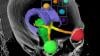
This is an example of the Mirada DLCExpert deep learning software that automatically identifies organs, segments and auto-contours them as the first step in creating radiation oncology treatment plans. This example of a segmented prostate computed tomography (CT) scan being used to plan radiotherapy was created without any human intervention. It was demonstrated at the American Association of Physicists in Medicine (AAPM) 2019 meeting.
This example shows OAR Space hydrogel (outlined in blue) injected to create space between the prostate and the rectum to prevent damage to that radiation sensitive structure. The gel is hard to identify on the CT scan because it looks like part of the rectum or prostate. But the softwares AI has been trained to identify it when present.
The DLCExpert software was cleared by the FDA in July 2018 and was first shown at ASTRO 2018. It automatically identifies anatomical structures and contours them to save staff time. The files created by the software are vendor neutral and can be imported into any vendor’s treatment planning system. Read more about this software.
Patient Positioning Radiation Therapy | August 21, 2019

This is a quick demonstration of the Varian Identify image-guided patient positioning system at the American Association of Physicists in Medicine (AAPM) 2019 meeting. It helps align patients on the radiotherapy system treatment table to match the position they were in when the computed tomography (CT) scan was created. This ensures the radiation beams are delivered according to the treatment plan and will not be aimed accidentally at health tissue. It uses real-time tracking of the surface of the patient's skin using three visible light emitters, so it does not add dose, such as when on-board X-ray imaging is used. The system compares the patients position to the treatment plan CT scan and color codes in red any areas that are not in the proper position. It also uses RFID tags on the table to help know the exact position of the patient.
The system can show the radiotherapist if the patient is no longer aligned with the plan and the therapist can manually stop the therapy. The vendor said in the future, they plan to integrate the system with Varian's therapy systems so treatment will be stopped automatically by the Identify system.
The system also uses a biometric scanner to ensure the correct plan is being used with the correct patient.
Treatment Planning | August 21, 2019

This is a lung cancer tumor radiotherapy treatment plan for the Accuray CyberKnife system demonstrated at the American Association of Physicists in Medicine (AAPM) 2019 meeting. The blue lines are the radiation beam lines that are shot from different positions to all intersect in the tumor to deliver the prescribed amount of radiation and prevent damage to surrounding healthy tissue. The beams also are planned around the critical structure organs near the target tumor to limit their dose. The organs are color coded to differentiate them on the treatment plan and to help with the estimated radiation dose each receives based on the plan. After the plan is optimized, it is fed into the radiotherapy treatment system computer to deliver the treatment once the patient is positioned on the treatment table exactly as they are in the CT scans used to create the plan.
Find more news and video from AAPM
Computed Tomography (CT) | August 21, 2019

This is a quick walk around of a mobile 32-slice computed tomography (CT) scanner used for surgery, brachytherapy and proton therapy on display by Mobius Imaging at the 2019 American Association Of Physicists in Medicine (AAPM) meeting. The system simply plugs into a standard wall outlet and all of the required hardware and software is built into the gantry. There is no need for an equipment closet, cabinet or server tower. The company said the CT system was created by some of the same developers who built the O-arm mobile CT system, but they said this CT scanner is much more compact.
Radiographic Fluoroscopy (RF) | August 09, 2019

Shimadzu displayed the FluoroSpeed X1 conventional radiographic fluoroscopy (RF) system at the Association for Medical Imaging Management (AHRA) 2019 meeting in July. The system was pending U.S. Food and Drug Administration (FDA) approval at AHRA, but received FDA 510(k) clearance in early August 2019.
The system features a 33-inch aperture, large enough to place a wheelchair inside. It can be rotated 90 degrees in either direction and the deck can be parked in any position, making it easier for patients to get on and off the 660-pound weight table. The FluoroSpeed X1 offers controls that are ergonomic for technologists, with duplicate controls on each side for either a left- or right-handed tech. The machine also has a large aperture to allow swallow studies.
The FluoroSpeed X1 comes equipped with a 17 x 17-inch dynamic digital X-ray detector (FPD) in the table bucky, allowing it to both be used for fluoroscopy as well as radiographic exams.
Read more about the FluoroSpeed X1: Shimadzu Medical Systems Receives FDA 510(k) for FluoroSpeed X1 RF System
Interact with a 360 photo of a Shimadzu FluoroSpeed X1 Fluoroscopy imaging system
CT Angiography (CTA) | August 07, 2019

This is a quick walk around of the new Siemens Somatom Go.top cardiovascular edition compact computed tomography (CT) scanner on display at the Society Of Cardiovascular Computed Tomography (SCCT) 2019 meeting in July. It is aimed at cardiology office based imaging and was released this past spring at the American College of Cardiology (ACC) meeting.
The system has removable tablets on each side of the scanner where the tech can adjust the machine, review scout scans and trigger the scanner. The idea is to improve workflow and allow the tech to remain at the bedside longer to be with the patient, rather tucked away in a remote control room using an intercom.
The entire system is built into the gantry seen here, so there is no need for extra equipment in a closet, cabinet or server tower.
It comes in a 128 slice configuration with 4 cm of anatomical coverage per rotation.
It uses the Stellar detector and tin filtration to eliminate low energy photons and help lower dose. It can be programmed to aid workflow by automatically removing bone, create cured planar reconstructions, lung CAD and other post-processing features so more time can be spent on reading scans. The scanner also comes with a HeartFlow FFR-CT starter pack.
Find more information on this system in these related articles:
New Cardiovascular CT Technology Entering the Market
New Technology Highlights on the ACC 2019 Exhibit Floor
CT Angiography (CTA) | August 07, 2019

This is a quick walk around of the GE Healthcare Cardiographe dedicated cardiac CT system on display at the Society Of Cardiovascular Computed Tomography (SCCT) 2019 meeting. It was designed specifically for cardiac imaging and so has a very compact footprint so it can be used in an office setting or small room. It offers a fast gantry rotation speed to freeze cardiac motion and has large enough anatomical coverage to view the scan the entire heart in one rotation.
One of these systems was recently installed at St. Paul’s Hospital in Vancouver, Canada, where they have an extensive structural heart program. Read more about this intall.
Find more information on this system in these related articles:
New Cardiovascular CT Technology Entering the Market
New Technology Highlights on the ACC 2019 Exhibit Floor
Radiology Business | August 02, 2019

Association for Medical Imaging Management (AHRA) President Chris Tomlinson, CRA, FAHRA, and President-elect Jacqui Rose, CRA, FAHRA, discuss some of the most important clinical topics at the 2019 AHRA Annual Meeting and how the association plans to help its members embrace technological change in the coming years. Among the main focuses at the meeting were clinical decision support (CDS), artificial intelligence (AI) and the use of data analytics to improve equipment and personnel performance.
Watch the VIDEO: Assessing Cardiovascular Risk in Ultra-endurance Athletes, an interview with Colorado State University graduate research assistant Nate Bachman at AHRA 2019.
Cardiac Imaging | July 30, 2019

Nate Bachman, graduate research assistant in the Human Cardiovascular Physiology Lab of the Dept. of Health and Exercise Science at Colorado State University, describes how he and fellow researchers used multiple types of cardiac imaging to evaluate the health of athletes who compete in endurance events lasting six hours or more, and what the results may suggest for future screening.
Watch the VIDEO: Key Topics for Radiology Administrators at AHRA 2019, an interview with AHRA President Chris Tomlinson, CRA, FAHRA, and President-elect Jacqui Rose, CRA, FAHRA.
Radiation Therapy | July 30, 2019

Pierre Qian, MBBS, cardiac electrophysiologist fellow, Brigham and Women's Hospital, explains how his facility is working with radiation oncology to use radio therapy to noninvasively ablate ventricular tachycardia (VT). He spoke on this topics during a joint electrophysiology session by the Heart Rhythm Society (HRS) and the Society of Cardiovascular Computed Tomography (SCCT) at the SCCT 2019 meeting.
Cardiac Imaging | July 30, 2019

Mark Ibrahim, M.D., FACC, assistant professor of medicine and radiology, associate program director, advanced cardiac imaging fellowship, University of Utah, explains what radiologists and cardiologists need to know what is needed from CT imaging prior to ablation procedures for atrial fibrillation (AF) and ventricular fibrillation (VF). He spoke at a joint session of the Heart Rhythm Society (HRS) and the Society of Cardiovascular Computed Tomography (SCCT) at the 2019 SCCT meeting.
Cardiac Imaging | July 30, 2019

Arthur Agatston, M.D., clinical professor of medicine, Florida International University, Herbert Wertheim College of Medicine, is the name-sake of the Agatston score used in CT calcium scoring. He explains the history of the scoring system from the early 1990s and the evolution of CT technology for cardiac imaging. The latest American Heart Association (AHA) 2018 cholesterol guidelines now include the use of CT calcium scoring, which was a big topic at the Society of Cardiovascular Computed Tomography (SCCT) 2019 meeting.
Related CT Calcium Scorining Content:
VIDEO: New Cholesterol Guidelines Support CT Calcium Scoring for Risk Assessment — Interview with Matthew Budoff, M.D.
CT Calcium Scoring Becoming a Key Risk Factor Assessment
ACC and AHA Release Updated Cholesterol Guidelines for 2018
VIDEO: CT Calcium Scoring to Screen For Who Should Take Statins — Interview with Matthew Budoff, M.D.
Find more SCCT news and videos
Computed Tomography (CT) | July 30, 2019

Cynthia McCollough, Ph.D., director of the Mayo Clinic CT Clinical Innovation Center, professor of medical physics and biomedical engineering and the 2019 president of the American Association of Physicists in Medicine (AAPM), shares her insights on the latest advances in computed tomography (CT) imaging technology. She spoke at the 2019 AAPM meeting. She also did an interview at AAPM on her president's theme for the 2019 meeting - VIDEO: Bridging Diversity in Medical Physics to Improve Patient Care.
Find more news and videos from AAPM.
Related CT Technology Content:
New CT Technology Entering the Market
VIDEO: Advances in Cardiac CT Imaging — Interview with David Bluemke, M.D.
Expanding Applications for Computed Tomography
VIDEO: Overview of Cardiac CT Trends and 2019 SCCT Meeting Highlights —Interview with Ron Blankstein, M.D., direct
VIDEO: 10 Tips to Improve Cardiac CT Imaging — Interview with Quynh Truong, M.D.
FFR-CT: Is It Radiology or Cardiology?
VIDEO: ITN Editor's Choice of the Most Innovative New Technology at RSNA 2018
VIDEO: Using Advanced CT to Enhance Radiation Therapy Planning — Interview with Carri Glide-Hurst, Ph.D.
VIDEO: Tips and Tricks to Aid Cardiac CT Technologist Workflow
VIDEO: ITN Editor's Choice of Most Innovative New Cardiac CT Technology at SCCT 2017
New Developments in Cardiovascular Computed Tomography at SCCT 2017
VIDEO: Role of Cardiac CT in Value-based Medicine — Leslee Shaw, Ph.D.
Advances in Cardiac Imaging Technologies at RSNA 2017
VIDEO: The Future of Cardiac CT in the Next Decade — Interview with Leslee Shaw, Ph.D.
VIDEO: What to Consider When Comparing 64-slice to Higher Slice CT Systems — Interview with Claudio Smuclovisky, M.D.
AAPM | July 29, 2019

Brent Parker, Ph.D., DABR, professor of radiation physics and medical physicist at MD Anderson Cancer Center, explains how the American Association of Physicists in Medicine (AAPM) is creating guidelines to better define the roles of non-physicist assistants. He said there is a lack of state regulatory oversight for medical physicists or their assistants, partly because there are no guidelines from the medical societies. AAPM has created a series of policy statements to better define these the roles and requirements for all of these positions. Parker said the goal is to give state regulators the the definitions needed to create oversight guidelines. He spoke on this topic in sessions at the AAPM 2019 meeting.
AAPM | July 29, 2019

Mahadevappa Mahesh, Ph.D., chief of medical physicist and professor of radiology and medical physics, Johns Hopkins University, Baltimore, and treasurer of the American Association of Physicists in Medicine (AAPM), explains some of the trends in medical physics and new features of the AAPM 2019 meeting.
Watch the related VIDEO: Bridging Diversity in Medical Physics to Improve Patient Care — Interview with AAPM President Cynthia McCollough, Ph.D., at the 2019 AAPM meeting.
Radiation Therapy | July 23, 2019

Paul Liu, Ph.D., post-doctoral research associate, Image X Institute at the University of Sydney, Australia, explains how his center is working on a low-cost radiation therapy system for the developing world. The Nano-X system will use a fixed linac gantry and rotate the patient around the beam. This would lighten the weight of the system, reduce the need for room shielding, and cut the number of moving parts to lower costs and ease maintenance. Liu spoke about the project in sessions at the American Association of Physicists in Medicine (AAPM) 2019 meeting.
AAPM | July 23, 2019

Cynthia McCollough, Ph.D., director of the Mayo Clinic Computed Tomography (CT) Clinical Innovation Center, professor of medical physics and biomedical engineering, and the 2019 president of the American Association of Physicists in Medicine (AAPM), explains the "building bridges" theme of the 2019 AAPM meeting.
This theme was the focus of her president’s address at the 2019 AAPM meeting. She spoke on the theme of diversity and how to break down the barriers between various minorities, male-female, religion, national origin, etc. She gave many photo examples of how we pigeon hole people into neat categories and that we often say we have equally in society, however her images showed recent images of big political summits where there are no women present, or they were the secretaries in the background. She said in medical practice, department administration and collaboration on projects, people need to be cognoscente of bias they have engrained by culture for which they may not even be aware.
She showed a slide of the AAPM membership makeup by generation and said members need to keep in mind the way each generation thinks and communicates varies by their generation's life experience and upbringing. McCollough said understanding these differences can help bridge perceived gaps in communication.
Brachytherapy Systems | July 23, 2019

Lior Arazi, Ph.D., assistant professor at Ben-Gurion University, Israel, explains the potential benefits of a new Radium-224 brachytherapy seed technology he is helping develop. The technology uses high-dose alpha particles to kill cancer cells, but has a very short tissue penetration, so it can be placed very close to critical structures without causing collateral damage to healthy tissue. He discussed this technology in sessions at the American Association of Physicists in Medicine (AAPM) 2019 meeting.
Radiation Oncology | July 22, 2019

Stephen Sorensen, Ph.D., DABR, chief of medical physics, St. Joseph's Hospital and Medical Center, Phoenix, Arizona, explains the first commercial use of the Zap-X stereotactic radio surgery (SRS) brain radiotherapy system. The system uses a capsule-like shield to surround the gantry and patient, eliminating the need for expensive room build outs requiring vaults. The goal of the system is to expand SRS brain therapy by making it easier and less expensive to acquire the treatment system. Sorensen spoke about this system in sessions at the American Association of Physicists in Medicine (AAPM) 2019 meeting.
Find more news and videos from AAPM.
Artificial Intelligence | July 22, 2019

Leigh Conroy, Ph.D., physics resident, University Health Network, Princess Margaret Cancer Center, Toronto, Canada, explains how her center is using machine learning to automate treatment plans. The center is one of the first to use the RayStation machine learning treatment planning system for radiation oncology. She spoke at the American Association of Physicists in Medicine (AAPM) 2019 meeting.
Learn more about how this technology works in the VIDEO: Editor's Choice of the Most Innovative Technologies at AAPM 2018.
Find more news and videos from AAPM.
CT Angiography (CTA) | July 19, 2019

Ron Blankstein, M.D., director of cardiac computed tomography, Brigham and Women's Hospital, and associate professor of medicine and radiology, Harvard Medical School, offers an overview of the recent trends in cardiac CT and some of the new highlights at the Society of Cardiovascular Computed Tomography (SCCT) 2019 meeting. He said key topics included integration of artificial intelligence into CT systems, the integration of CT calcium scoring into the 2018 American Heart Association (AHA) cholesterol management guidelines, structural heart assessments for transcatheter valve and left atrial appendage (LAA) occlusion, and sessions with paertner societies that explain the roll of CT in interventional cardiology and electrophysiology.
Computed Tomography (CT) | July 19, 2019

Quynh Truong, M.D., MPH, associate professor of radiology and medicine at Weill Cornell and director of cardiac CT, NewYork Presbyterian Hospital, offers 10 tips to help improve image quality for cardiovascular computed tomography (CTA) exams. She spoke on this topic at the Society of Cardiovascular Computed Tomography (SCCT) 2019 meeting.
Artificial Intelligence | July 12, 2019

Khan Siddiqui, M.D., founder and CEO of HOPPR, discusses the economic advantages and costs presented by artificial intelligence (AI) applications in radiology, as well as potential strategies for healthcare providers looking to add AI to their armamentarium, at the 2019 Society of Imaging Informatics in Medicine (SIIM) annual meeting.
Digital Pathology | July 11, 2019

Toby Cornish, M.D., Ph.D., associate professor and medical director of informatics at the University of Colorado School of Medicine, explains how the subspecialty of digital pathology has evolved in recent years, the benefits of integrating pathology and radiology, and how artificial intelligence (AI) may smooth the transition, at the 2019 Society of Imaging Informatics in Medicine (SIIM) annual meeting.
Enterprise Imaging | July 09, 2019

ITN Associate Editor Jeff Zagoudis speaks with Don Dennison, healthcare IT consultant and Chris Roth, M.D., associate professor of radiology, vice chair, information technology and clinical informatics, and director of imaging informatics strategy at Duke University Medical Center, about how to find the right people to deploy a successful enterprise imaging strategy.
Watch part 1 of the interview at the 2019 Society for Imaging Informatics in Medicine (SIIM) conference.
Enterprise Imaging | July 08, 2019

ITN Associate Editor Jeff Zagoudis speaks with Don Dennison, healthcare IT consultant and Chris Roth, M.D., associate professor of radiology, vice chair, information technology and clinical informatics, and director of imaging informatics strategy at Duke University Medical Center, about how to find the right people to deploy a successful enterprise imaging strategy.
Artificial Intelligence | July 03, 2019

Sudhen Desai, M.D., FSIR, interventional radiologist at Texas Children's Hospital, editor of IR Quarterly for the Society of Interventional Radiology (SIR) and on the Board of Directors for the Society of Physician Entrepreneurs, explained how artificial intelligence (AI) can assist in pediatric imaging and the pitfalls of training AI systems. He spoke at the 2019 Radiology AIMed conference.
Deep learning algorithms require large amounts of patient case data to train the systems to read medical images automatically without human intervention. However, in pediatrics, there are often much lower numbers of normal and abnormal scans that can be used compared to vast amounts of adult exams available. This makes it difficult to train systems, so AI developers are coming up with innovative new ways to train their software. Compounding issues with training pediatric imaging AI is that the normal ranges change very quickly for young children due to their rapid development. He explained what is normal for a 2-year-old may not be normal for a 5-year-old.
Desai and other pediatric physicians who spoke at the conference said AI could have a big impact on pediatric imaging where there are not enough specialists for the increasing image volumes.
Related content:
VIDEO: Implementation of Artificial Intelligence Tools in Radiology Practice — Interview with Lawrence Tanenbaum, M.D.
VIDEO: AI That Second Reads Radiology Reports and Deals With Incidental Findings — Interview with Nina Kottler, M.D.
Technology Report: Artificial Intelligence at RSNA 2018
VIDEO: Implementation of Artificial Intelligence Tools in Radiology Practice
Interventional Radiology | June 26, 2019

Kevin Seals, M.D., University of California San Francisco (UCSF) Health, interventional radiology fellow, is working on a research project using smart speakers such as the Amazon Echo and Google Home to create a new method for accessing information on device technologies in real time in the interventional radiology (IR) lab. Operators can use the conversational voice interface to retrieve information without breaking sterile scrub. The technology uses using natural language processing (NLP) and machine learning to rapidly provide information about device sizing and compatibility in IR.
Seals spoke at the 2019 Radiology AIMed conference in Chicago in June.
Related content:
Atrium Health Debuts Amazon Alexa Skill to Help Patients Access Medical Care
Smart Speaker Technology Harnessed for Hospital Medical Treatments
Artificial Intelligence | June 26, 2019

Nina Kottler, M.D., vice president of clinical operations, Radiology Partners, explains how the company developed its own artificial intelligence (AI) application to check the accuracy of radiologists' reports as they dictated and to follow-up automatically on incidental findings. She spoke at the 2019 Radiology AIMed conference.
She said the AI sits on top of Radiology Partners' natural language processing application and will immediately flag any comments that do not appear to make sense or might need clarification. Instead of forcing radiologists to change their workflow to create structured reports, Kottler said this AI software helps adapt the existing workflow to make the reports more consistent and able to be data mined later.
Related content:
VIDEO: Implementation of Artificial Intelligence Tools in Radiology Practice — Interview with Lawrence Tanenbaum, M.D.
VIDEO: Artificial Intelligence May Assist in Pediatric Imaging — Interview with Sudhen Desai, M.D.
Technology Report: Artificial Intelligence at RSNA 2018
VIDEO: Implementation of Artificial Intelligence Tools in Radiology Practice
Artificial Intelligence | June 21, 2019

Lawrence Tanenbaum, M.D., Radnet vice president and chief technology officer, discusses some of the artificial intelligence (AI) products in radiology that are now commercially available and how AI developments will impact PET, MRI and CT imaging. He spoke at the 2019 Radiology AI-Med conference.
He said AI is helping medical imaging in the following areas:
• Identify urgent findings and flagging these on the top of worklists.
• Computer aided detection capabilities that go beyond the traditional to improve efficiency, boost diagnosis and highlight unexpected findings.
• Improving diagnostic image reconstruction
• Tools to enhance the speed, resolution, radiation dose and overall quality of advanced imaging.
Learn more about what AI tools vendors are developing to help medical imaging in the following links:
Technology Report: Artificial Intelligence at RSNA 2018
VIDEO: Editor’s Choice of the Most Innovative New Artificial Intelligence Technologies at RSNA 2018
VIDEO: Artificial Intelligence May Assist in Pediatric Imaging — Interview with Sudhen Desai, M.D.
VIDEO: Editor's Choice of the Most Innovative New Technology at RSNA 2018
VIDEO: AI That Second Reads Radiology Reports and Deals With Incidental Findings — Interview with Nina Kottler, M.D.
VIDEO: RSNA Post-game Report on Artificial Intelligence
VIDEO: AI, Analytics and Informatics: The Future is Here — Interview with Michael Recht, M.D.
Radiation Therapy | May 21, 2019

This is a walk through of the ViewRay MRIdian MRI-guided radiotherapy system installed at Henry Ford Medical Center — Cottage. The system has been in service for about two years. The walk through shows the entry way into the vault and the warnings about entering an MRI environment. Anyone who enters the treatment room must undergo a metal detector scan first to make sure all ferrous metal is removed.
Read more about the center's first year experience with the system.