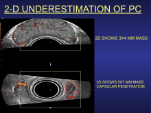
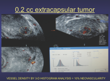
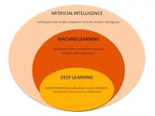
The use of smart algorithms has the potential to make healthcare more efficient. Sarah Eskreis-Winkler, M.D., presented data that such an algorithm — trained using deep learning (DL), a type of artificial intelligence (AI) — can reliably identify breast tumors in magnetic resonance (MR) images. In doing so, the algorithm has the potential to make radiology more efficient.


Pragmatism from cybersecurity to enterprise imaging was in vogue at the 2019 meeting of the Society of Imaging Informatics in Medicine (SIIM). Not unexpectedly, artificial intelligence accounted for much discussion amid telltale cracks in its hype.


Xifin announced the launch of the next evolution of its laboratory information system (LIS), Xifin LIS 6. The expanded platform features enhancements that support high-complexity, high-volume labs, as well as new integrated capabilities available through strategic partners, including artificial intelligence (AI)-powered digital pathology workflow and genomic data interpretation, automated prior authorization and test utilization decision support.Xifin announced the launch of the next evolution of its laboratory information system (LIS), Xifin LIS 6. The expanded platform features enhancements that support high-complexity, high-volume labs, as well as new integrated capabilities available through strategic partners, including artificial intelligence (AI)-powered digital pathology workflow and genomic data interpretation, automated prior authorization and test utilization decision support.

A new clinical guideline from the American Society for Radiation Oncology (ASTRO) provides recommendations on the use of radiation therapy to treat patients diagnosed with pancreatic cancer. Recommendations include when radiation treatments are appropriate, as well as the optimal dosing, timing and fractionation for these treatments. The guideline, which also outlines strategies to prevent and mitigate common side effects of pancreatic radiation therapy, is published online in Practical Radiation Oncology, the clinical practice journal of ASTRO.


In molecular radiotherapy (MRT) treatment of the thyroid, existing single photon emission computed tomography (SPECT) imaging systems are unable to accurately measure radiation absorbed by patients during and after treatment. As a consequence, only limited information regarding the success of radiotherapy treatment has been available. 


September 5, 2019 — An ahead-of-print article published in the December issue of the American Journal of Roentgenology (AJR) provides a primer of gender affirmation surgical therapies encountered in diagnostic imaging.[1] The article defines normal post-surgical anatomy and describes select complications using a multidisciplinary, multimodality approach. 

Heidelberg University Hospital, Germany, has treated its first patient using Monte Carlo photon dose planning in RayStation. The decision to go clinical was made after a thorough validation of the functionality. The first patient, who has cervical cancer involving the lymph nodes, was recently treated.


The global rise in chronic disease has significantly increased demand for diagnostic imaging procedures, and in turn, contrast media to enhance those images. More than 30 million imaging procedures are performed each year in the U.S., and 60 percent of contrast imaging agents sold are iodinated contrast agents.1 Although considered safe for the general population, iodinated contrast agents may place patients at an increased risk for nephrotoxicity, especially those with certain risk factors undergoing cardiac interventional procedures.


Extracapsular extension (ECE) is a major clinical indicator for brachytherapy or external beam treatment of prostate cancer. The prostatic capsule is a thin layer of compressed fibrous tissue and ultrasound imaging depicts the pericapsular fat as a strongly echogenic structure while magnetic resonance imaging (MRI) demonstrates a dark envelope. The capsule tends to be incomplete around the apex accounting for greater extraprostatic spread from apical tumors.


Iridium Cancer Network consists of all seven hospitals in the Antwerp region, closely collaborating regarding radiation oncology. The network acts as a single radiation therapy department, making it the largest in Belgium with its annual 5,300 patients. Iridium was first to use the RayCare* oncology information system (OIS) clinically, only two months after it was released from RaySearch.

Subscribe Now