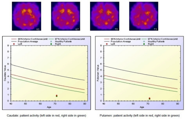
A 71-year-old male, previously submitted to high-dose chemotherapy due to advanced small cell lung cancer (SCLC). One year after the end of chemotherapy, he started experiencing cognitive impairment, mainly consisting of deficits in executive function and processing speed. He was submitted to 123I-FP-CIT SPECT which showed bilaterally severely reduced tracer uptake in the overall striatum, as demonstrated by axial slices (upper row) and by semiquantitative analysis (lower row) carried out through a dedicated software (Basal Ganglia Matching Tools 2).
Image created by Luca Filippi, Department of Nuclear Medicine, “Santa Maria Goretti” Hospital, Latina, Italy.
March 29, 2023 — A newly published literature review sheds light on how nuclear medicine brain imaging can help evaluate the biological changes that cause chemotherapy-related cognitive impairment (CRCI), commonly known as chemo-brain. Armed with this information, patients can understand better the changes in their cognitive status during and after treatment. This summary of findings was published ahead-of-print by The Journal of Nuclear Medicine.
CRCI describes a clinical condition characterized by memory and concentration impairment, difficulties with information processing and executive functioning, and mood and anxiety disorders. While CRCI has been widely investigated from a clinical perspective, little is known about the underlying biological mechanisms that cause chemo-brain.
“Nuclear medicine techniques can be used to investigate different physiopathological phenomena related to CRCI, such as cortical metabolism, dopamine transporter integrity, and neuroinflammation, with specific imaging probes,” said Agostino Chiaravalloti, MD, PhD, professor of nuclear medicine and nuclear medicine physician in the Department of Biomedicine and Prevention at University Tor Vergata in Rome, Italy. “However, nuclear medicine tests are not commonly considered in the work-up of patients with CRCI-related manifestations.”
To understand the current landscape of nuclear medicine and molecular imaging for chemo-brain, researchers undertook an extensive literature review. Following the PRISMA guidelines for literature searches, the researchers identified 22 relevant studies on two topics: 1) the effects of the most commonly used chemotherapy drugs on cognitive function and 2) the results of SPECT and PET examinations of CRCI. The findings confirmed the impact of chemotherapy drugs on cognitive function, such as impaired executive function, anxiety and trouble sleeping. They also highlighted the utility of various SPECT and PET imaging techniques to visualize glucose consumption, blood flow, or expression of receptors, all of which may play a role in CRCI.
In this context, nuclear medicine offers several instruments for the detailed evaluation of the physiopathological processes that underlie CRCI. “The findings presented could lead to a better understanding of the potential role of molecular imaging in the assessment of subtle changes in the brain after treatment and, possibly, in the monitoring of brain functions in patients treated with chemotherapy,” stated Chiaravalloti.
This study was made available online in February 2023.
The authors of “Functional imaging of chemo-brain: usefulness of nuclear medicine in the fog coming after cancer” include Agostino Chiaravalloti, Department of Biomedicine and Prevention, University of Rome Tor Vergata, Rome, Italy, and IRCCS Neuromed, Pozzilli, Italy; Lucca Filippi, Nuclear Medicine Section, "Santa Maria Goretti" Hospital, Latina, Italy; Marco Pagani, Institute of Cognitive Sciences and Technologies, Consiglio Nazionale Delle Ricerche (CNR), Rome, Italy, and Department of Medical Radiation Physics and Nuclear Medicine, Karolinska Hospital, Stockholm, Sweden; and Orazio Schillaci, Department of Biomedicine and Prevention, University of Rome Tor Vergata, Rome, Italy.
For more information: www.snmmi.org


 November 18, 2025


