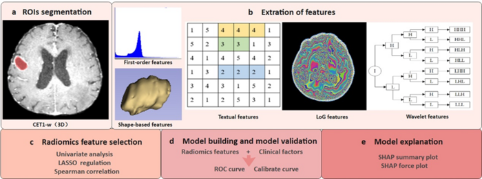
The workflow of radiomics. Image courtesy of Yixin Wang
June 24, 2022 — Recently, a collaborated research team led by Prof. LI Hai and Hongzhi Wang from Hefei Institutes of Physical Science of Chinese Academy of Sciences (CAS) proposed an interpretable radiomic model for predicting radiotherapy treatment response in patients with brain metastases.
The results were published in European Radiology.
Radiomics refers to extracting high-throughput radiomic features from medical images to assist clinical decision-making. These radiomic features can reflect the biological information of tumors, which cannot be obtained directly through conventional image interpretation. Therefore, machine learning-based methods can rely on in-depth data mining to obtain additional knowledge about tumor heterogeneity. Currently, there is no accurate prediction model of radiotherapy treatment response for patients with brain metastases in clinical practice.
In this research, the team proposed an interpretable radiomic model for predicting radiotherapy treatment response in patients with brain metastases by combining radiomics and SHAP methods to solve this clinical problem.
Yixin Wang, the first author of the paper, explained how they finished the whole process. At first, the research team extracted the radiomic features from the magnetic resonance imaging (MRI) images of patients with brain metastases before radiotherapy. Then they used the machine learning method to build the radiomic model. In the end, they explained the model using the game theory-based SHAP, which is helpful for the formulation of precise radiotherapy for patients with brain metastases.
The model had good performance, and the prediction results in the external validation group also showed that the model can be generalizability. At the same time, the SHAP method could realize the interpretability and visualization of the model and avoid the "black box" effect of traditional machine learning algorithms, which was beneficial for clinicians to understand the model and promote the use of the model.
This work was supported by the Key Research and Development Program of Anhui Province, the Collaborative Innovation Cultivation Fund of Hefei, Big Science Center of CAS, and the Key Clinical Cultivation Specialty of Hefei Cancer Hospital of CAS.
For more information: https://english.hf.cas.cn/
Related Brain Metastases Content:
PET Imaging Adds Valuable Information to Brain Metastasis Monitoring
ASTRO Issues Clinical Guideline on Radiation Therapy for Brain Metastases


 December 04, 2025


