23-Year-Old Man with Body Mass Index of 25.1. Left: UDFF—operator places crossbar at liver capsule, with sample ROI fixed 1.5 cm deep from crossbar, to ensure measurement obtained sufficiently deep to liver capsule. Overall UDFF was 3%. Right: Median MRI PDFF from three acquisitions was 3%, demonstrating agreement.
June 7, 2022 — According to ARRS’ American Journal of Roentgenology (AJR), ultrasound-derived fat fraction (UDFF) is strongly associated with MRI proton-density fat fraction (PDFF) and provides high sensitivity for detecting hepatic steatosis.
Noting that a reduced number of measurements is sufficient for determining overall UDFF (i.e., median value of the median of the five measurements for each of three acquisitions), “a UDFF cutoff of >5% provides high AUC and sensitivity, albeit low specificity, for detection of MRI PDFF ≥5.5%,” clarified corresponding author Andrew T. Trout of Cincinnati Children’s Hospital Medical Center in Ohio.
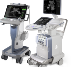
Trout and team’s cross-sectional study registered 56 overweight and obese adolescents and adults (≥16 years) who underwent investigational ultrasound (ACUSON Sequoia; deep abdominal transducer) and MRI examinations of the liver during a single visit from August to October 2020. Ultrasound examinations included three UDFF acquisitions of five measurements each. MRI examinations included three PDFF acquisitions with calculation of an overall median PDFF.
Ultimately, UDFF was positively associated with MRI PDFF (rho=0.82 [95% 0.71-0.89]; p<.001), with mean bias between methods of 4.0%. In addition, UDFF had an AUC of 0.90 (95% CI: 0.79-0.96, p<.001) for identifying an MRI PDFF ≥5.5%, with a cutoff value of >5% having 94.1% sensitivity and 63.6% specificity.
“In clinical practice, the authors of this AJR article concluded, “three measurements are likely sufficient for determining UDFF.”
For more information: www.arrs.org


 July 31, 2024


