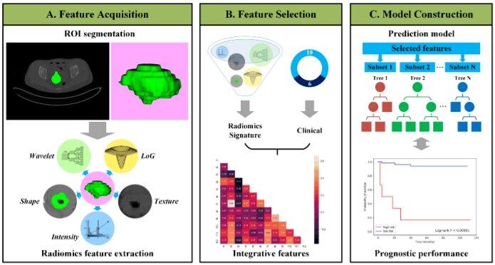
Fig. 1. Outline of the workflow from data input, feature extraction, selection, and model construction. Credit: DOI: 10.3233/CBM-210201
March 2, 2022 — Artificial intelligence (AI), deep learning (DL), and machine learning (ML) have transformed many industries and areas of science. Now, these tools are being applied to address the challenges of cancer biomarker discovery, where the analysis of vast amounts of imaging and molecular data is beyond the ability of traditional statistical analyses and tools. In a special issue of Cancer Biomarkers, researchers propose various approaches and explore some of the unique challenges of using AI, DL, and ML to improve the accuracy and predictive power of biomarkers for cancer and other diseases.
“The biomarker field is blessed with a plethora of imaging and molecular-based data, and at the same time, plagued with so much data that no one individual can comprehend it all,” explained Guest Editor Karin Rodland, PhD, Pacific Northwest National Laboratory, Richland; and Oregon Health and Science University, Portland, OR, USA. “AI offers a solution to that problem, and it has the potential to uncover novel interactions that more accurately reflect the biology of cancer and other diseases.”
Promising applications of AI, DL, and ML presented in this issue include identifying early-stage cancers, inferring the site of the specific cancer, aiding in the assignment of appropriate therapeutic options for each patient, characterizing the tumor microenvironment, and predicting the response to immunotherapy.
A comprehensive overview of the literature regarding the use of AI approaches to identify biomarkers for ovarian and pancreatic cancer illustrates underlying principles and looks at the gaps and challenges that face the field as a whole. Ovarian and pancreatic cancers are rare, but lethal because they lack early symptoms and detection. Lead investigator Juergen A. Klenk, PhD, Biomedical Data Science Lab, Deloitte Consulting LLP, Arlington, VA, USA, and colleagues describe studies using AI and ML to analyze images for the early detection of disease, and models that can be built to predict likely outcomes for the patient. Some of the challenges, such as the difficulty of gathering large enough datasets, are discussed.
“Algorithms develop biases and produce prejudiced responses when the data they are trained on are non-representative or incomplete,” Dr. Klenk said. The investigators suggest that the development of larger and more diverse image databases for rare cancers across institutions, standardized reporting methods, and easier-to-understand interfaces that increase user trust are needed to make a true impact on biomarker discovery.
Lead investigator Debiao Li, PhD, Biomedical Imaging Research Institute, Cedars-Sinai Medical Center, Los Angeles, CA, USA, and colleagues developed a model to identify individuals at risk for pancreatic ductal adenocarcinoma (PDAC). PDAC is associated with many preconditional abnormalities that can be visible on a computerized tomography (CT) scan, but these are difficult to comprehend by visual assessment. In their study, the investigators used CT scans from patients with confirmed PDAC and CT scans from the same patients who had had a CT scan six months to three years before diagnosis to identify a set of CT features that were potentially predictive of PDAC. The model was 86% accurate in classifying the patients and the healthy controls, using the identified CT features.
“The challenge of AI for the advancement of pancreatic cancer research is the scarcity of data due to low prevalence. The purpose of this proof-of concept model Is to encourage researchers to establish a larger dataset for extensive training and validation of the model,” said Dr. Li.
Radiomics is an emerging field where features are extracted from medical imaging using various techniques. Radiomic features can quantify tumor intensity, shape, and heterogeneity and have been applied to oncologic detection, diagnosis, therapeutic response, and prognosis. Lead investigators Shaoli Song, PhD, Shanghai Medical College and Fudan University, Shanghai, China, and Lisheng Wang, PhD, Shanghai Jiao Tong University, Shanghai, China, and colleagues combined radiomic data from preoperative positron emission tomography (PET) and CT images in patients with early stage uterine cervical squamous cell carcinoma. They used algorithms to develop a prognostic signature capable of predicting disease-free survival.
“This model could provide more accurate information about potential relapse and metastasis, and could be helpful in decision-making,” they observed.
Other papers in the special issue focus on the development of new computational tools to facilitate the application of AI to biomarker identification; the use of whole cell imaging and immunofluorescence to identify immune features in pancreatic tumors to provide prognostic information; the use of microRNAs and applied machine learning to identify a miRNA profile associated with gastrointestinal stromal tumors; and the use of hierarchical clustering of combined multi-omic datasets to identify an antitumor immune signature in patients with colon cancer.
Dr. Rodland added that the articles in this special issue are only a small sampling of the various approaches to using AI, DL, and ML in biomarker research. “There is a continuing urgent need for more effective strategies for improving the early detection of cancers. Cutting-edge AI systems have been shown to improve sensitivity and specificity in the interpretation of both imaging and non-imaging data for breast, lung, prostate, and cervical cancers,” she stated.
For more information: www.iospresscom


 December 04, 2025


