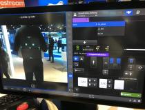
This gallery includes photos of medical imaging technologies from across the expo floor at the Radiological Society of North America (RSNA) 2021 annual meeting Nov. 28 - Dec. 2.
If you enjoy this content, please share it with a colleague
-
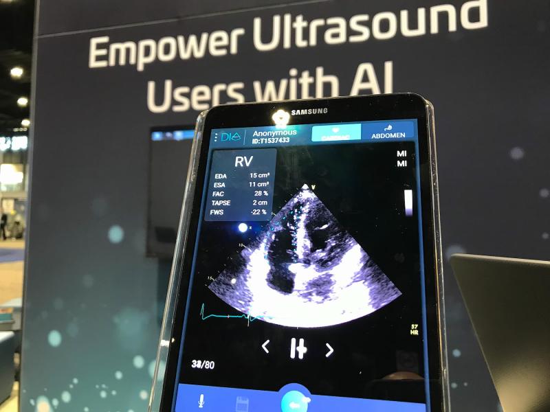
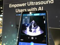
Dia provides artificial intelligence for cardiac ultrasound to automate ejection fraction and other functions. In the point-of-care ultrasound market the company can help automate cardiac analysis with AI and get consistent results, even with less experienced sonographers. #RSNA2021 #RSNA #RSNA21 #POCUS
-
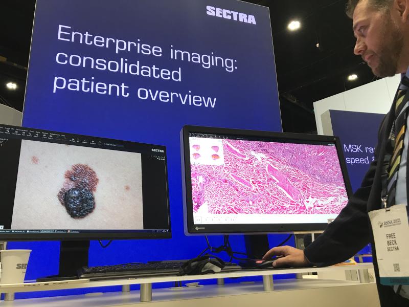
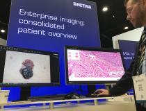
Newer enterprise imaging platforms enable storage of all types of non-DICOM data and images. This example from Sectra shows its system can pull up digital pathology slides and available light JPEG images of a suspected skin cancer. Hospitals are looking at enterprise imaging solutions to enable better images and report sharing across various departments beyond radiology and cardiology and into the EMR. #RSNA #pathology #digitalpathology #RSNA21
-

-
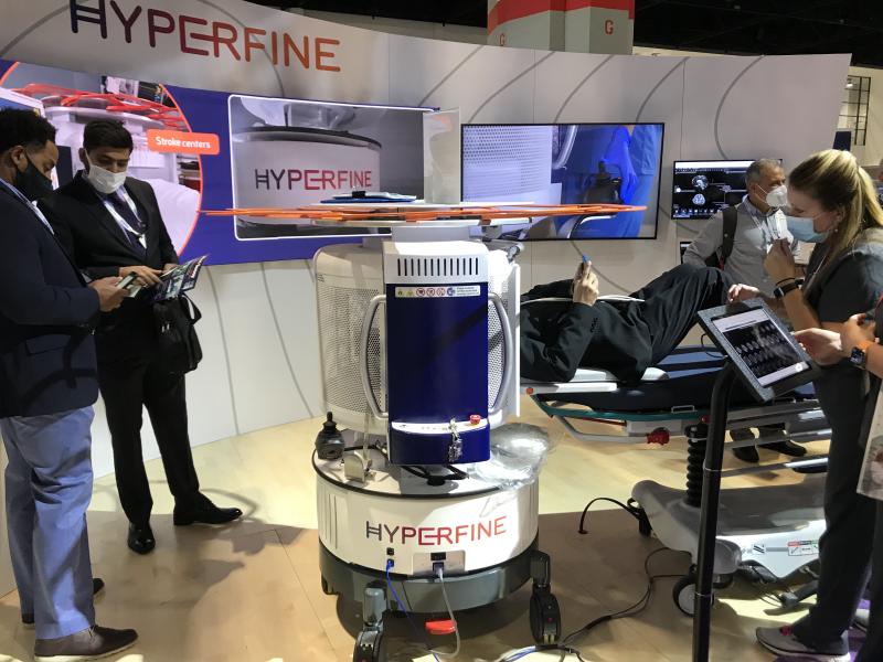
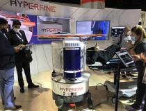
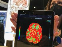
The first person to even have an MRI performed on the RSNA show floor, while other attendees wait their turn to get a brain scan in the first 15 minutes of the show floor opening on the first day of RSNA 2021. Hyperfine displayed its FDA-cleared Swoop head MRI system, and was among one of the hottest stops of the expo floor this year.
-
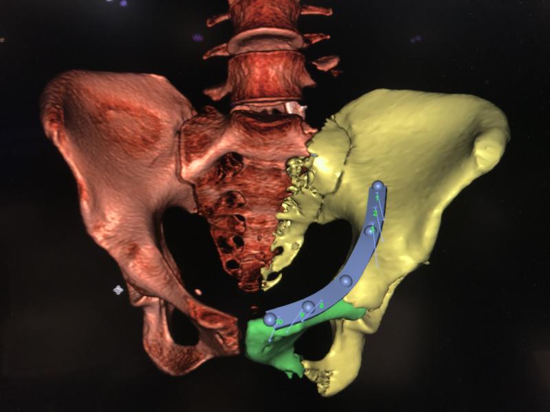
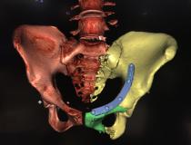
Fractured hip CT image reconstruction with orthopedic repair planning shown on the Sectra enterprise imaging system. #RSNA21 #RSNA2021 #RSNA
-
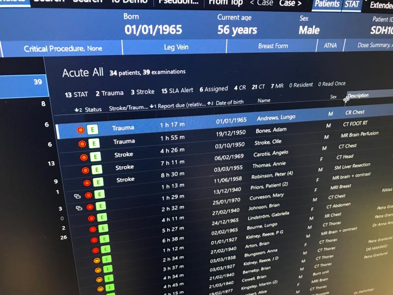
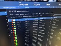
Example of an automated workflow orchestration DICOM worklist on the Sectra enterprise imaging system. The system prevents cherry picking of studies by assigning them to radiologists based on load balancing protocols, the specialties of each reader, the level of read priority and parameters of service level agreements (SLAs) with various hospitals. Workflow orchestration is offered by several vendors as a way to improve radiology workflow and to incorporate rules for priority reads and contract obligations.
-
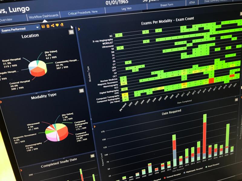
Example of an analytics capabilities of the Sectra workflow orchestration system. It can give a clear picture of what radiologists are doing in the system, reads they perform and business metrics for various kinds of exam reads and parameters set in service level agreements (SLAs) with various hospitals.
-
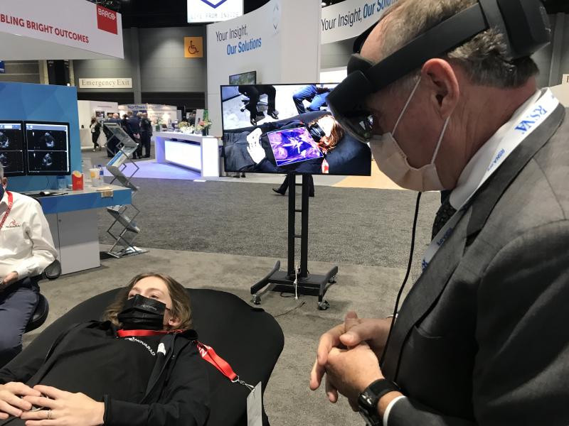
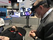
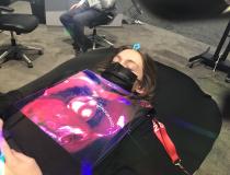
Novarad demonstrated augmented reality (AR) technology that can fuse cardiac MRI with a live patient on the table and tools for needle guidance for a pericardiocentesis.
-
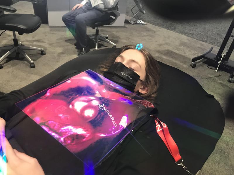
Novarad demonstrated augmented reality (AR) technology that can fuse cardiac MRI with a live patient on the table and tools for needle guidance for a pericardiocentesis.
-
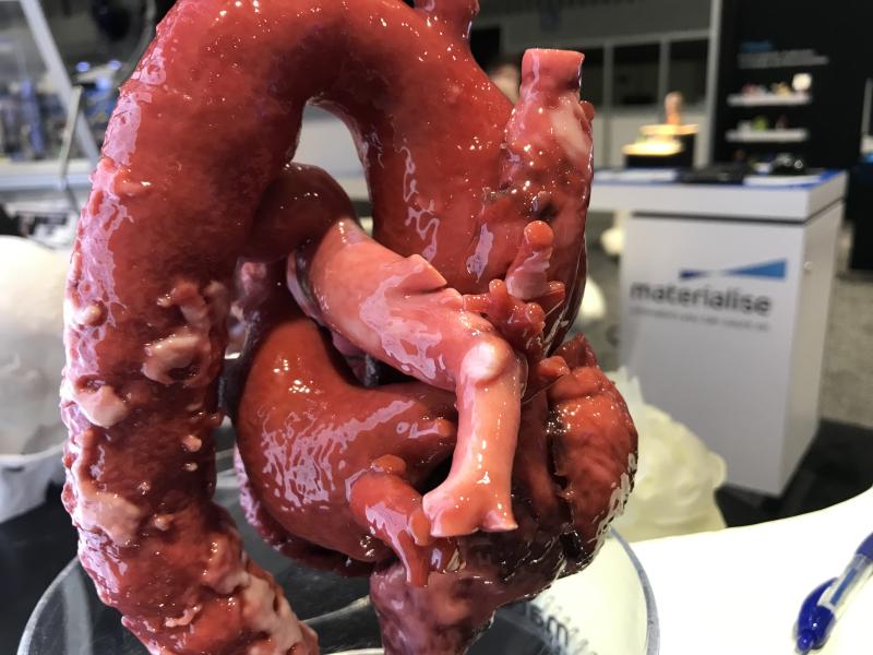
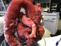
Example of life-like 3D printed cardiac and aortic anatomy available from Materialise. The vendor's software is FDA-cleared for use to print anatomy from CT or MRI studies that will be the exact size as the patient's actual anatomy. This can be used for planning and practicing complex surgical or interventional procedures and the model can aid navigation during procedures.
-
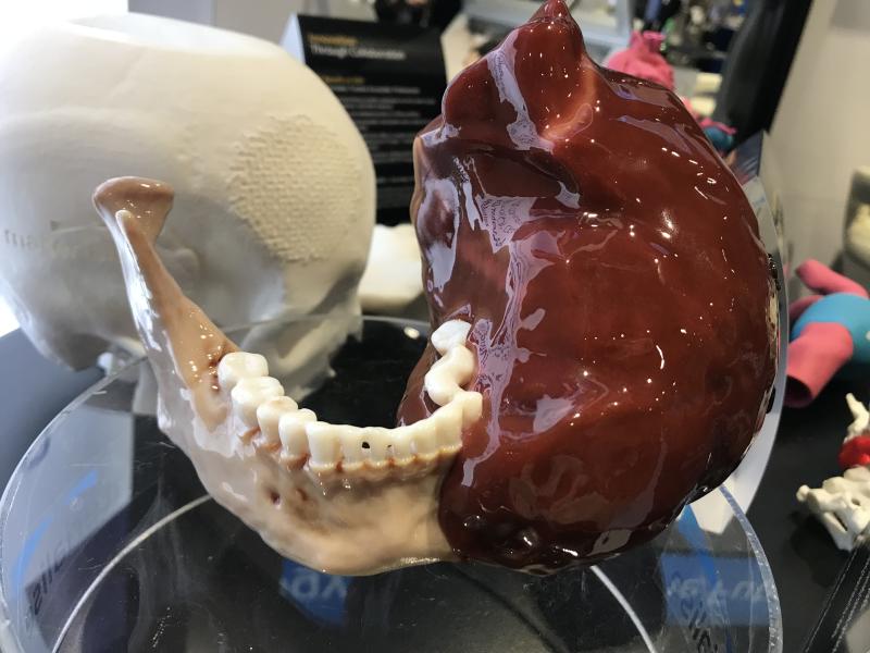
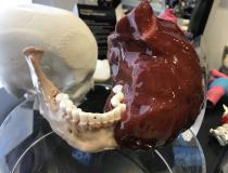
Example of life-like 3D printed tumor that is incorporated into a patient's jaw bone from Materialise. The vendor's software is FDA-cleared for use to print anatomy from CT or MRI studies that will be the exact size as the patient's actual anatomy. This can be used for planning and practicing complex surgical or interventional procedures and the model can aid navigation during procedures.
-
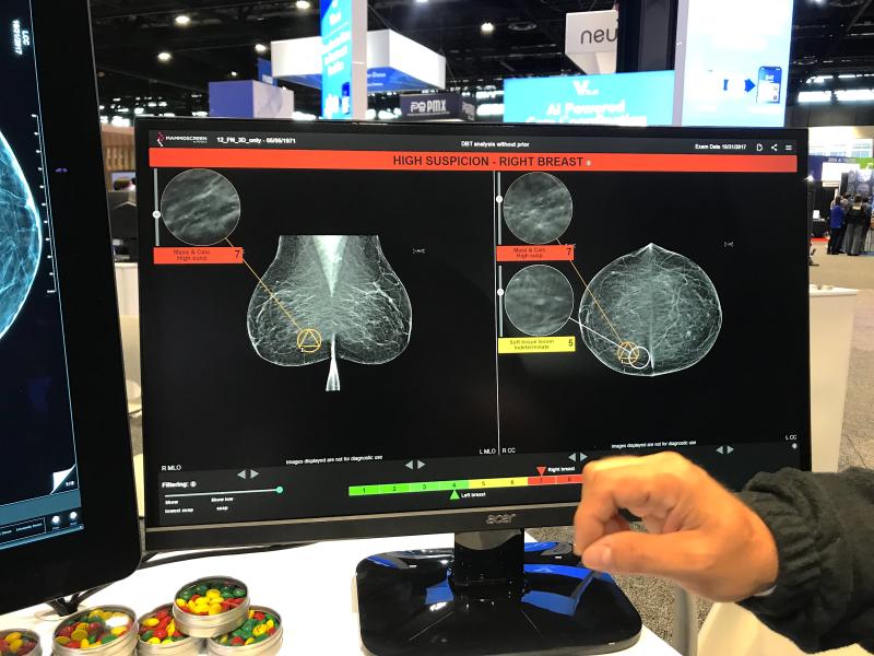
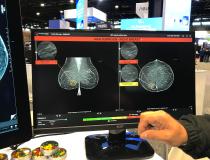
MammoScreen's artificial intelligence for mammography. The system shows the AI analysis on a different screen for the radiologist rather than embedding it on the mammograms. This allows the radiologist to review it after they read the study to serve as a second set of eyes.
-
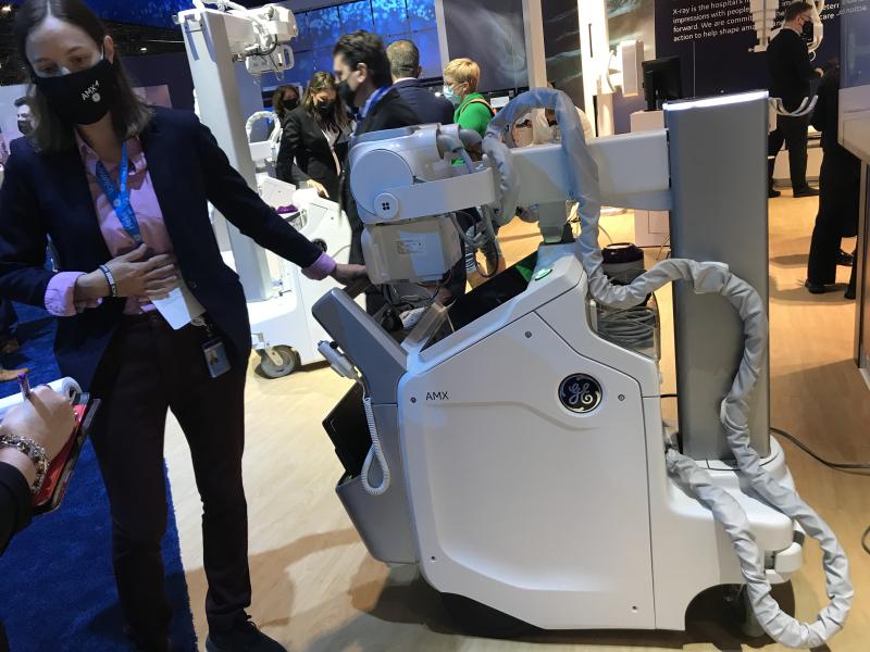
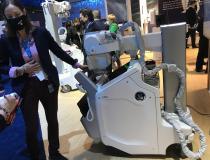
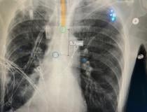
The GE Healthcare AMX mobile DR X-ray system. It includes an artificial intelligence feature to detect both misplaced endotracheal tubes and pneumothorax of the machine, so the attending physician can immediately be notified.
-
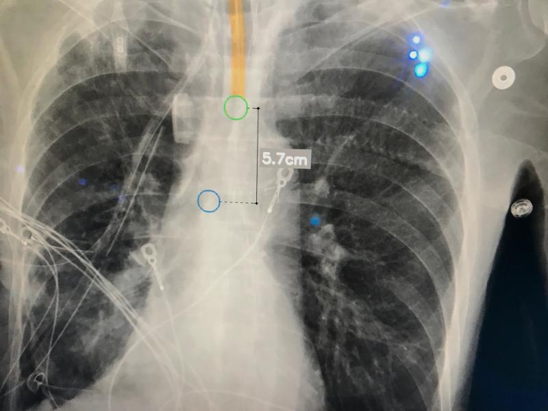
The GE Healthcare AMX mobile DR X-ray system includes an artificial intelligence feature to detect misplaced endotracheal tubes so the attending physician can immediately be notified.
-
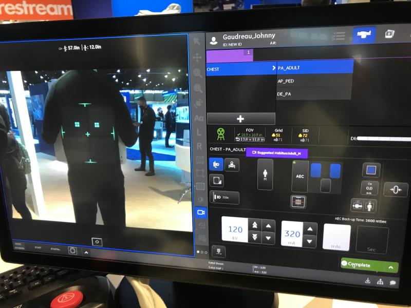
GE Healthcare's new Definium Tempo fixed, overhead tube suspension digital X-ray system is designed to be a "personal assistant" to radiologists and technologists to improve workflow. This includes a lot of automation to reducing workflow burdens. In patient setup, this includes Auto Positioning, Auto Centering and Auto Tracking, to automate the positioning of system components to maximize overall ergonomic operation for the technologist and improve overall patient experience.
-
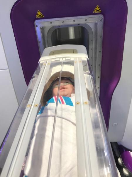
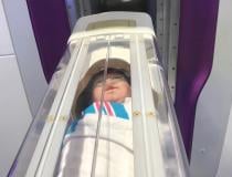
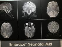
This is the Aspect Imaging Embrace 1T neonate MRI system is a dedicated NICU system. It is self-shielded so it does not require a special imaging room and allows babies to be imaged in the NICU instead of being transported to radiology. The capsule the neonate is placed in is temperature controlled and the noise level is about 65 decibels. It gained FDA in 2017 and also has CE mark. It is installed in 3 U.S. hospitals and one in Israel.
-
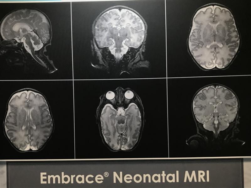
This is the Aspect Imaging Embrace 1T neonate MRI system is a dedicated NICU system. It is self-shielded so it does not require a special imaging room and allows babies to be imaged in the NICU instead of being transported to radiology. The capsule the neonate is placed in is temperature controlled and the noise level is about 65 decibels. It gained FDA in 2017 and also has CE mark. It is installed in 3 U.S. hospitals and one in Israel.
-
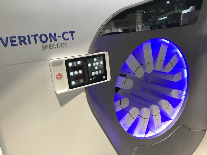
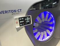
The Spectrum Dynamics Veriton SPECT-CT system at RSNA 2021. It with comes in 16, 64 or 128 slice CT configurations. It has 12 SPECT detector robotic arms that automatically move toward the patient and use a sensor to stop a few millimeters from the skin to optimize photon counts and SPECT image quality. It also uses more sensitive CZT digital detectors, which allows either faster scan times, or use of only half the radiotracer dose of analog detector scans.
-
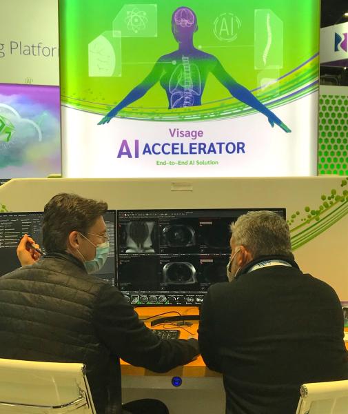
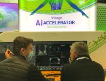
Visage Imaging demonstration on the floor of its artificial intelligence applications.
-
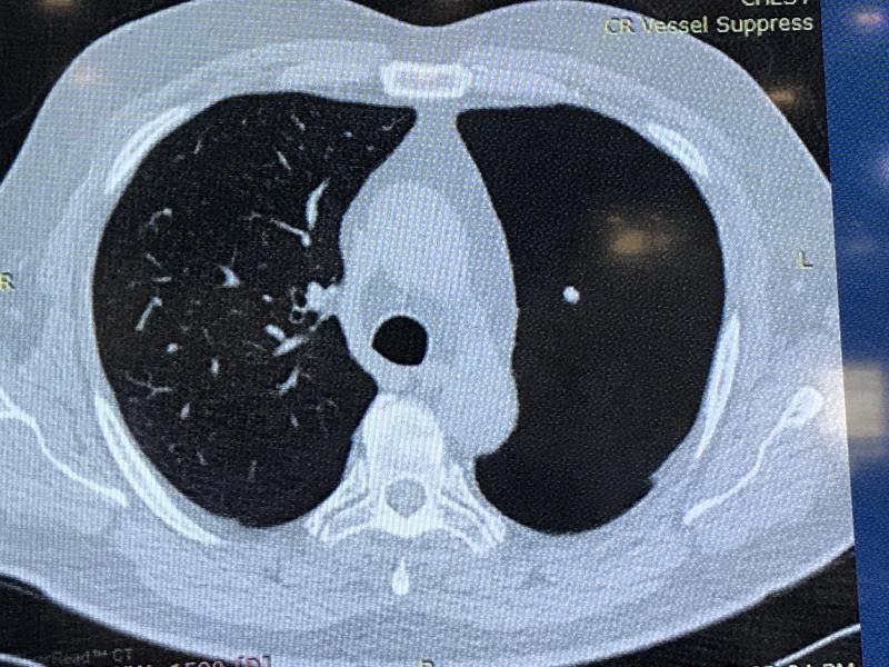
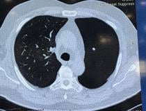
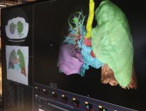
Example of Riverain's pulmonary artery vessel suppression artificial intelligence application for CT. It can automatically remove pulmonary vessels the system to just show lung nodules that the AI had determined are not cross sectional views of vessels.
-
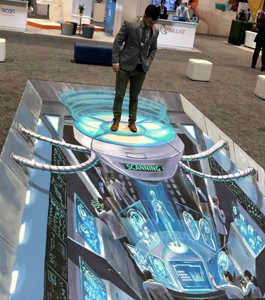
Artwork that invites attendees to interact with it for photos on the RSNA expo floor.
-
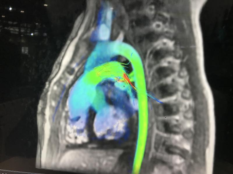
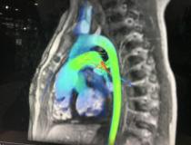
AI-enabled cardiac MRI flow analysis from Arterys.
-
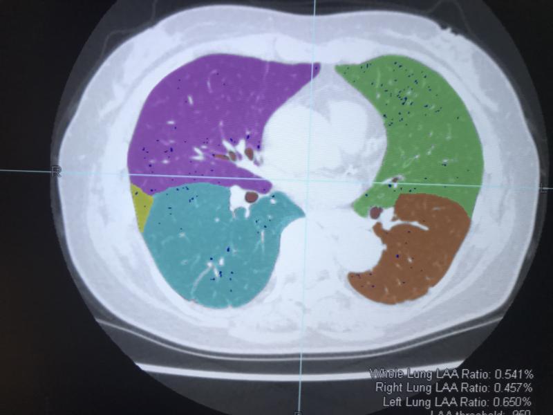
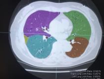
Ziosoft introduced a new lung nodule analysis application on its advanced visualization platform.
-
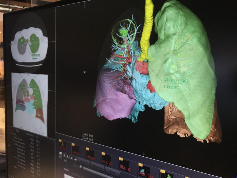
Ziosoft introduced a new lung nodule analysis application on its advanced visualization platform.
-
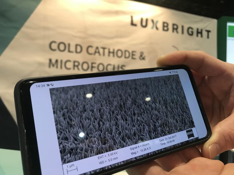
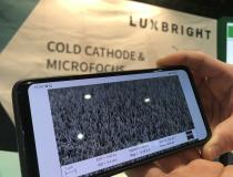
X-ray tube vendor Luxbright is developing a cold-cathode X-ray tube made from zinc-oxide nano crystals. The vendor showed what the crystals look like under a microscope on their phone. They hope to start testing the new tube in 2022. Several companies are working on cold-cathode technology. While working tubes have been made and even commercialized, issue with them appears to be longevity, as they wear out quickly compared to conventional hot-cathode tube technology used for the past 130 years. Luxbright is
-
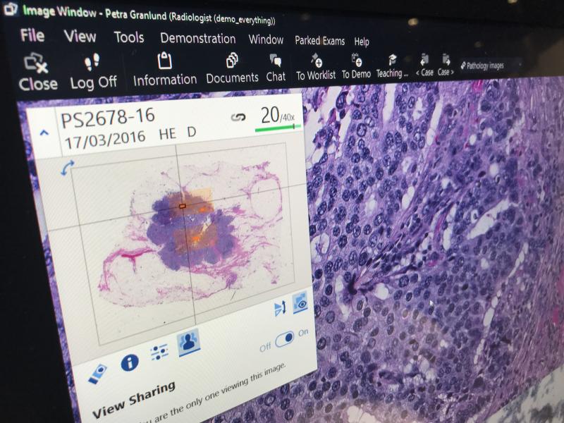
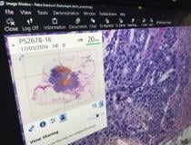
An example of a digital pathology microscope slide and a magnified image of the cells beneath it displayed by Sectra. Digital pathology is becoming a bigger part of RSNA as hospitals look at enterprise imaging system to use as a single image repository for all their image-heavy departments.
-
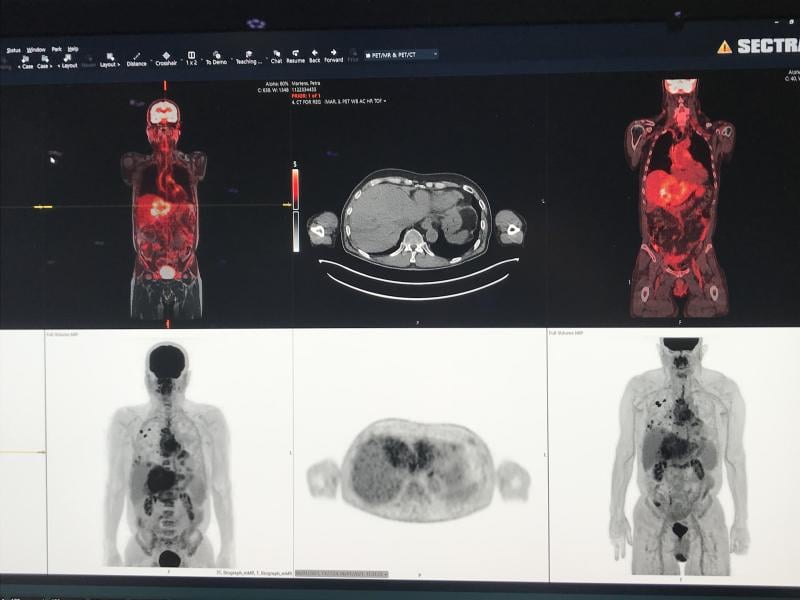
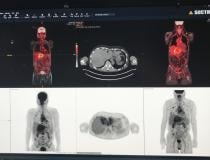
Example of nuclear imaging PET-CT showing a liver cancer patient with multiple metastases. Shown as part of a demo on the Sectra enterprise imaging system.
-
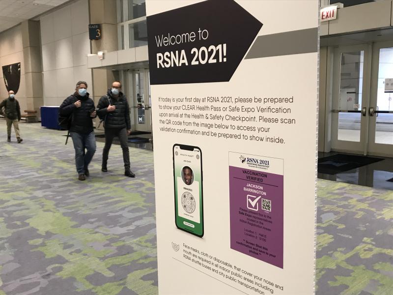
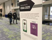
RSNA 2021 required all attendees to show proof of vaccination and required wearing a mask at all times.
-
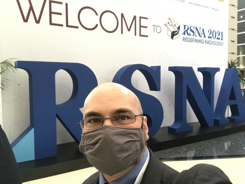
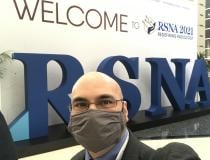
ITN and DAIC Editor Dave Fornell at RSNA 2021.
-
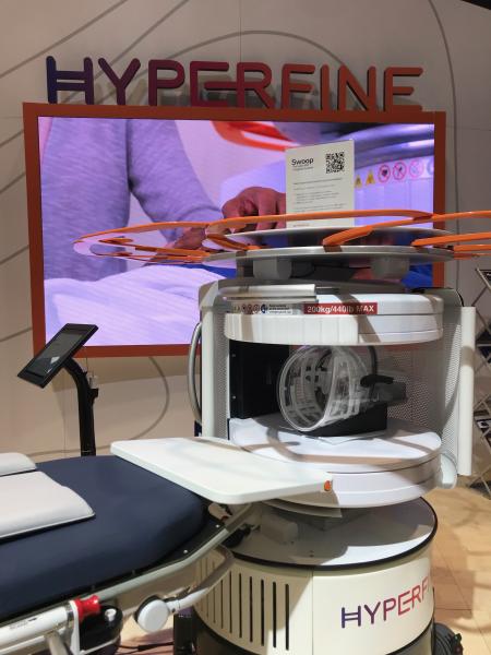
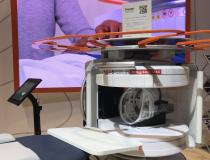
The Hyperfine Swoop portable MRI system on display at RSNA 2021. The company performed live MRI brain scans on anyone who wanted to be scanned on the show floor. The system is well below 1T and is self shielded so it was safe top operate on the floor. The system has FDA clearance and is designed for use in patient rooms and in the emergency department.
-
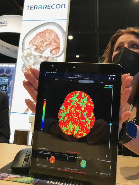
A demonstration of how advanced visualization reconstructions on the TeraRecon iNtuition system can easily be viewed and shared on a mobile device. The company has been very involved in AI the past few years at RSNA, but refocused attention on its third-party advanced visualization platform this year.
-
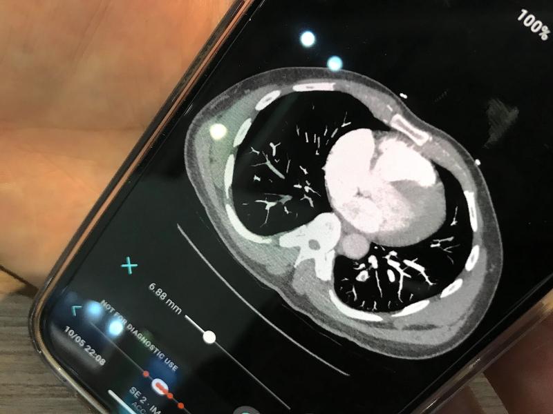
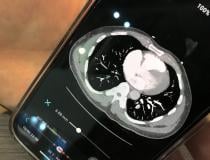
Aidoc's artificial intelligence pulmonary embolism response team (PERT) activation app showing how the CT scan can be viewed on a smartphone. The orange dots at the bottom of the page mark key slices were the AI detected pulmonary emboli.
-
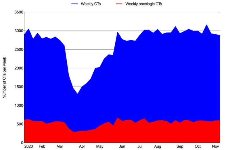
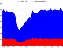
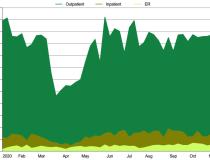
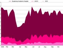
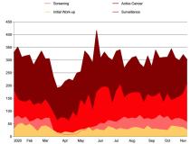
Figure 1. COVID weekly volumes of all CTs and oncologic CTs from January 5, 2020, to November 14, 2020.
-

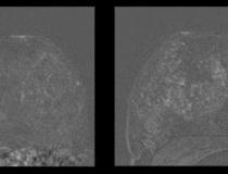
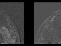
Left image shows breast MRI in 41-year-old patient without IUD. Right image shows increased parenchymal enhancement in the same patient 27 months after IUD placement. Image courtesy of RSNA
-
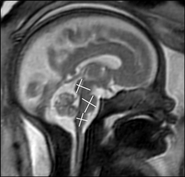
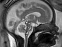
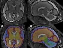
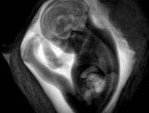
This images shows an MRI of fetal brain development. Image courtesy of RSNA
-
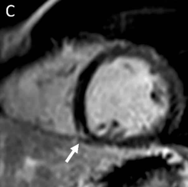
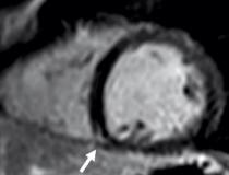
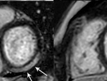
MRI shows late gadolinium enhancement (LGE) at the right ventricular attachment (arrow) of a 19-year-old male. Image courtesy of RSNA
-
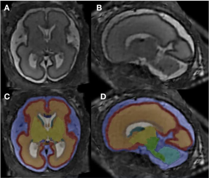
MRI super-resolution reconstruction and atlas-based tissue segmentation. Image courtesy of RSNA
-
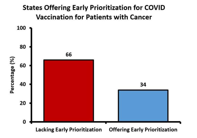
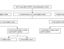
This chart shows the proportion of U.S. states offering equivalent early prioritization for COVID vaccination for patients with cancer and patients over the age of 65 as recommended by the Centers for Disease Control and Prevention.
-
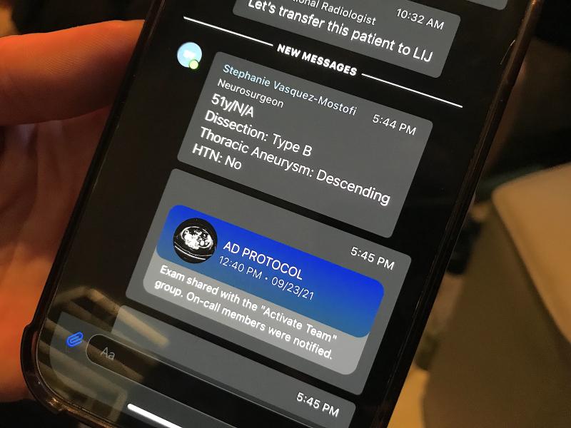
Viz.AI showed an example of an aortic dissection care team alert system. It uses AI to detect a type A or B dissection and immediately alerts the care team before the images are even loaded into the PACS. The AI sends a link to the imaging and this images shows the dissection on the CT.
-
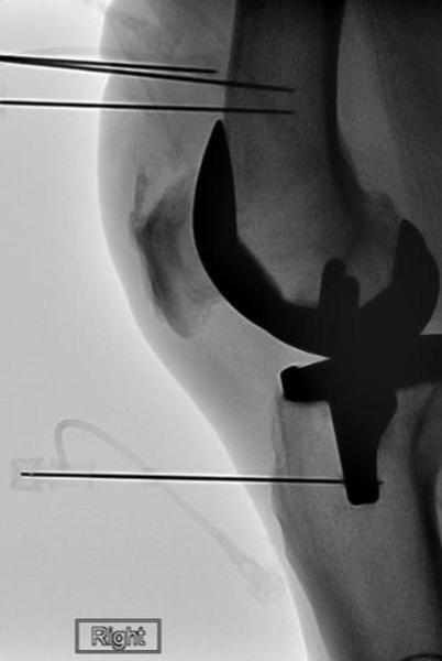
Radiofrequency ablation electrode is placed into the introducer needle after the placement of the introducer needle. Positioning was verified with imaging. Image courtesy of RSNA
-
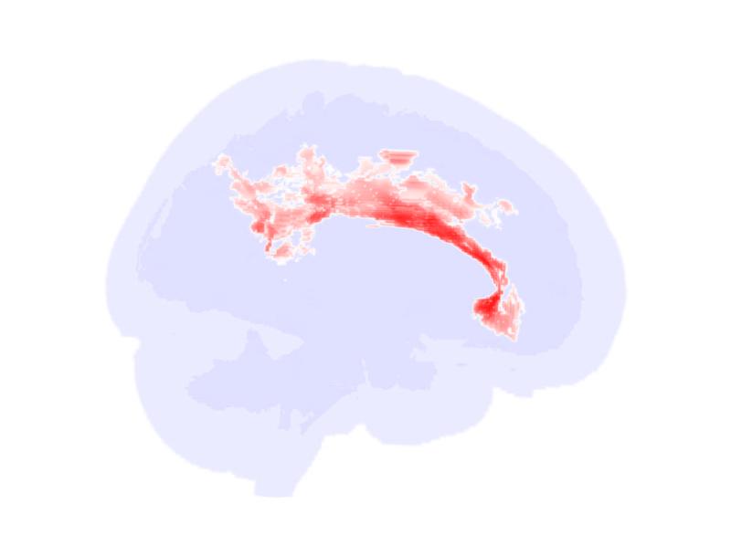
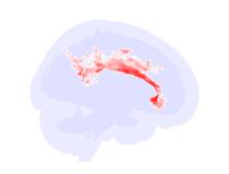
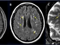
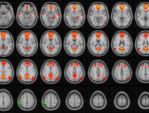
Significant alterations in the brain’s white matter in adolescents with ASD. Image courtesy of RSNA
-
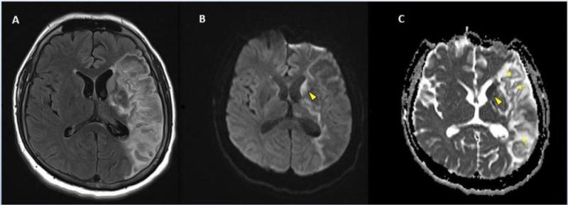
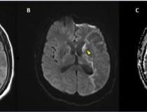
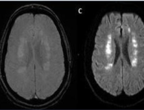
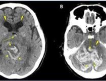
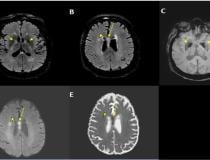
Stroke seen in a 41-year-old male patient with COVID-19 infection. Image courtesy of RSNA
-
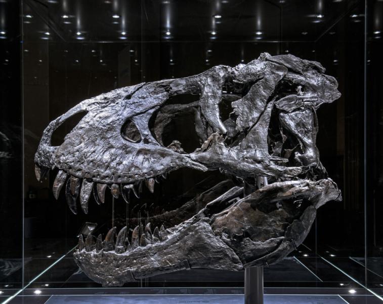
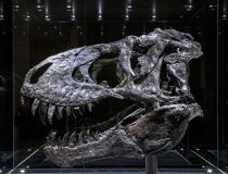
The “Tristan Otto” Tyrannosaurus rex skull that was examined by researchers, and presented in a study during RSNA21.
-

-

Acute anterior cerebral artery/middle cerebral artery watershed infarction seen in a 47-year-old male patient who presented with COVID-19 pneumonia. Image courtesy of RSNA
-

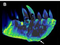
CT reconstructions of the tooth-bearing part of the left dentary. (A) Reconstruction of the conventional CT images in lateral view showing well-preserved anatomical structures such as the replacement teeth. Image courtesy of RSNA
-
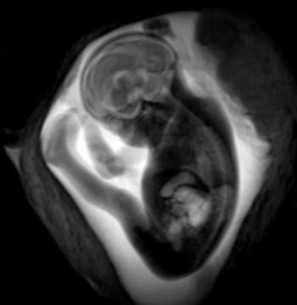
COVID-19 of mild to moderate severity in pregnant women appears to have no effect on the brain of the developing fetus, according to a study being presented today at the annual meeting of the Radiological Society of North America (RSNA).
-

Left image shows 45-year-old patient with IUD in place. Right image shows breast MRI of the same patient 32 months after IUD removal. Image courtesy of RSNA
-
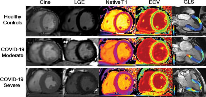
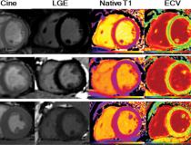
LGE images, native T1 maps, extracellular volume maps, and global longitudinal strain (GLS) show myocardial abnormalities in adults recovering from moderate and severe COVID-19. Image courtesy of RSNA
-
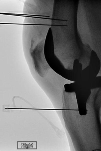
Radiofrequency ablation electrode is placed into the introducer needle after the placement of the introducer needle. Positioning was verified with imaging. Image courtesy of RSNA
-

Schematic demonstrating online availability of COVID-19 vaccine eligibility information for cancer patients at the state level during February 2021. Centers for Disease Control and Prevention
-
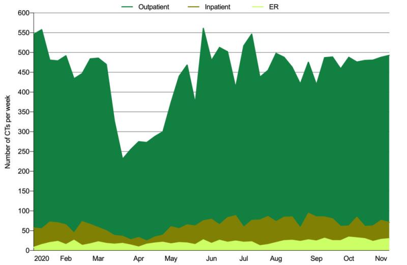
This chart shows weekly oncologic CT volumes by imaging indication from January 5, 2020, to November 14, 2020. Courtesy of RSNA
-
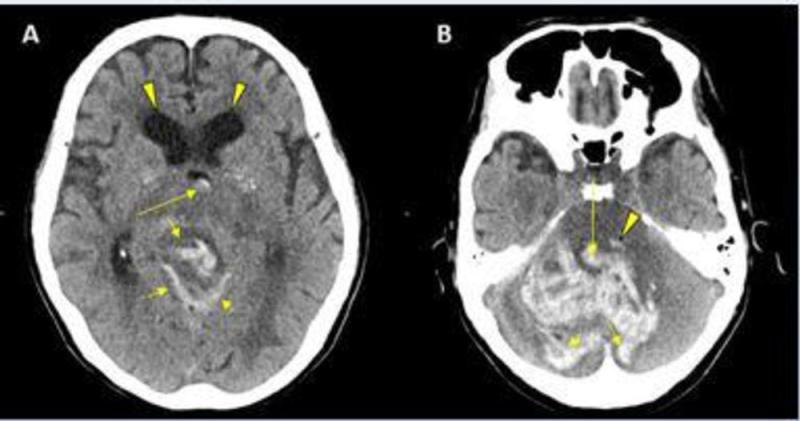
CT showing hemorrhage in a 68-year-old male patient with COVID-19 infection. Image courtesy of RSNA
-
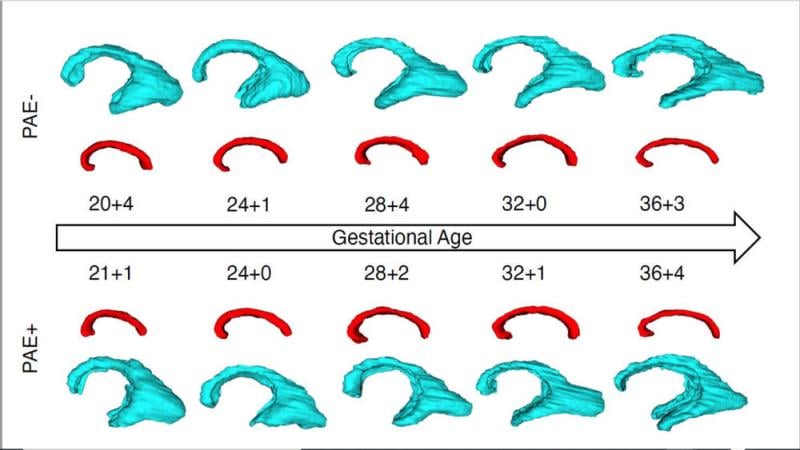
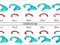
Longitudinal growth trajectories of the periventricular zone (blue) and the corpus callosum (red) in fetuses with (PAE+) and without (PAE-) prenatal alcohol exposure. Image courtesy of RSNA
-
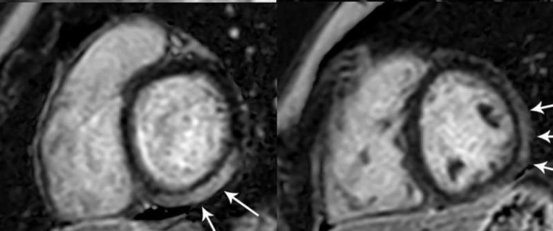
MRI shows prominent LGE involving more than 50% of the myocardium in the basal inferolateral and inferior segments of the left ventricle. Image courtesy of RSNA
-
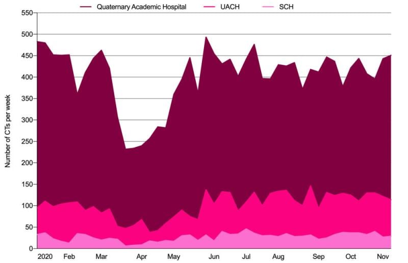
This chart outlinesoncologic CTs performed per week by hospital type. Courtesy of RSNA
-
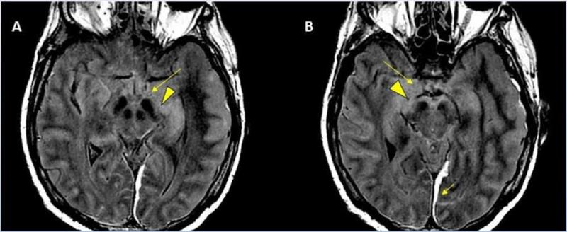
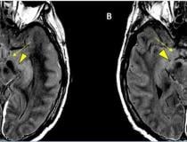
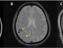
A 62-year-old male with a past medical history of hypertension presenting with seizures. Image courtesy of RSNA
-
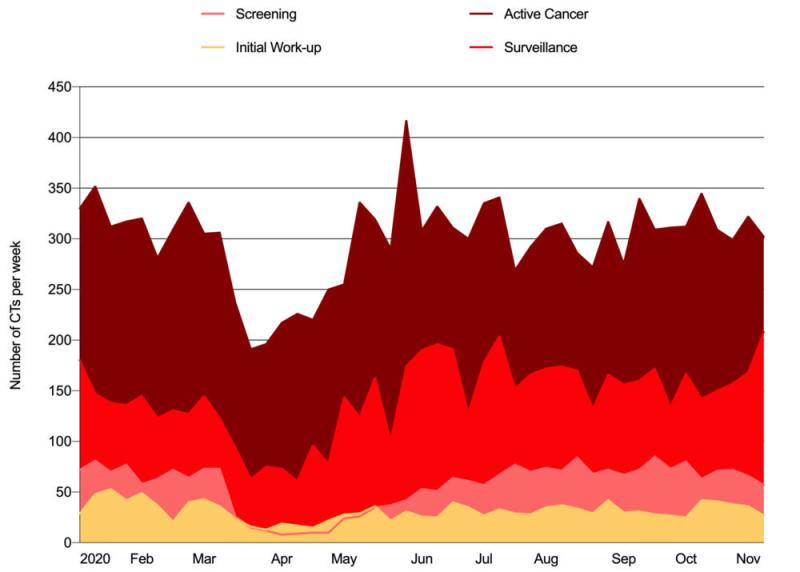
Weekly oncologic CT volume by care setting from January 5, 2020, to November 14, 2020. Courtesy of RSNA
-
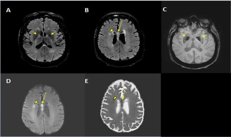
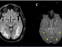
A 56-year-old male patient with diabetes and hypertension who presented with complaints of confusion. Courtesy of RSNA
-
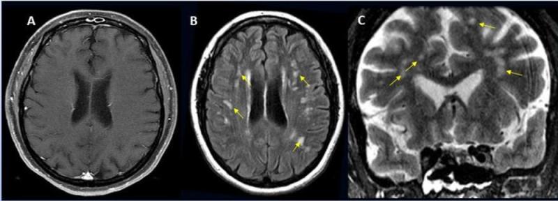
A 49-year-old female with past medical history of mitral valve disease and tricuspid valve regurgitation who developed headache followed by cough and fever presented to the ER with right upper eyelid ptosis (drooping). Courtesy of RSNA
-

Hemorrhage seen in 56-year-old female with COVID-19 infection and no other significant past medical history. Courtesy of RSNA
-

A 65-year-old male smoker, presented with acute hypoxic respiratory failure secondary to COVID-19 pneumonia, requiring intubation. Courtesy of RSNA
-

MedChat's livechat with text message lets you meet your patients where they are.
-
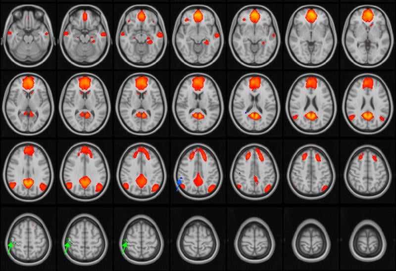
An MRI image of the brain. Courtesy of RSNA
-
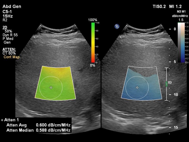
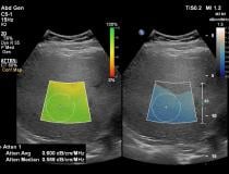
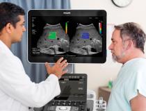
Philips Healthcare Epiq Ultrasound GI Liver
-
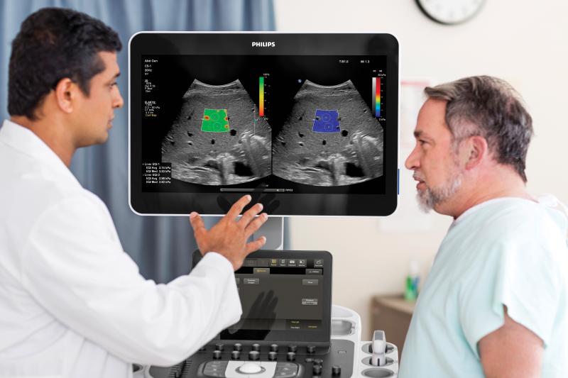
The Philips liver fat quantification tool.
-
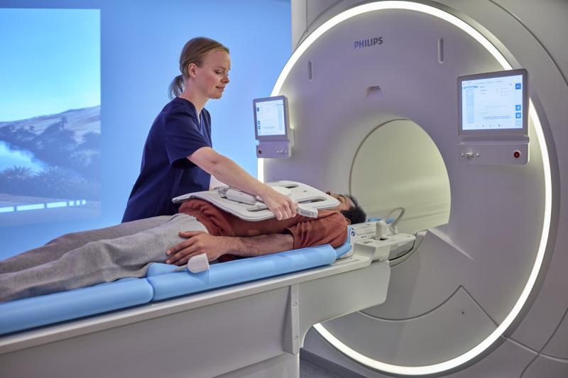
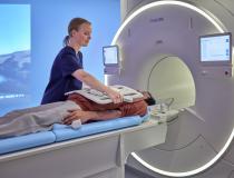
The Philips mr5300 in use.
-
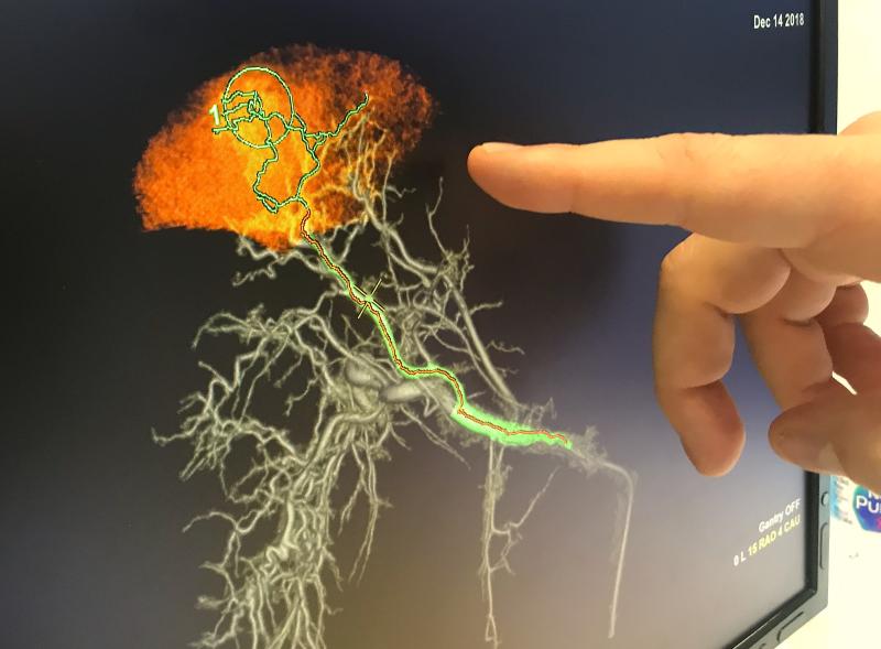
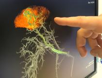
GE Healthcare's new version of its Liver Assist embolization guidance software at RSNA 2021. It uses a rotational angiography sweep to create a cone-beam CT 3D image reconstruction of the liver vessels and the target lesion. The new version also shows the parenchyma (functional area of the anatomy) for a tumor. This can help differentiated the tumor tissue from healthy liver tissue when choosing an embolization site.
-

This was the first in-person RSNA since 2019 due to the pandemic. Many attendees used the opportunity to get together with distant colleagues and old friends. This was a gathering at the Hyatt after the conference was winding down for the day at RSNA.
-
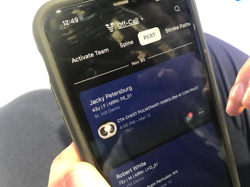
The Viz.AI pulmonary embolism response team (PERT) app that uses AI to detect PE on images before they go into PACS. The AI automatically alerts the PERT team members on their cell phones and provides a mobile platform to message, review images and other patient data.
-
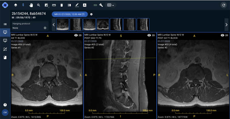
Sirona Medical, a software company founded on a deep understanding of both the practice and business of radiology, announced the launch of its cloud-native radiology operating system (RadOS) at the 2021 Annual Meeting for Radiological Society of North America (RSNA). The unified workspace platform was developed based on the principle that technology, and AI in particular, should augment the intelligence of the physician.

