April 29, 2019 — A new report by Arthur Olch, Ph.D., highlights use of specialized software at Children’s Hospital Los Angeles (CHLA) that could advance treatment accuracy of radiation oncology for pediatric cancer patients.
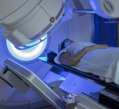
During radiation therapy, patient position must be stable from session to session to ensure radiation beams are properly targeting the tumor. For this reason, X-ray images are taken before each treatment. Radiation therapists can use this information to reorient the patient so that the position is exactly the same each time. Doctors at CHLA are taking this already rigorous process one step further. From early in its development Olch, a radiation physicist, has been evaluating the use of new software to advance quality assurance in radiation therapy. In a recent publication, he highlights the use of this technological advance to aid in the treatment of pediatric cancers.
Radiation therapy uses a beam of targeted X-rays that kill cancer cells over the course of treatment. After the beam passes through the patient, it is captured on an imaging panel. Olch and his team make use of the information carried by these beams, called exit images, using the automated software. These images contain important information about the exact dose being delivered to the tumor and surrounding tissues and can be compared to the planned doses. Up to 20 images might be generated per treatment session. With treatments occurring every day for several weeks, this makes for an unwieldy amount of data to manually process. Now, radiation oncology staff have a tool that will do this in seconds. The program automates not only image capture but also analysis.
Analyzing these images provides new information that allows further fine tuning of the radiation beams and patient position from session to session. This, said Olch, gives radiation oncologists more information that can be used to account for anatomy changes in real time. "If a patient gains or loses weight, their dimensions change" he said. "Likewise, as the tumor shrinks, radiation beams need to take a different trajectory."
Adjustments are routinely made as a standard of care, but by utilizing the latest technological advances, CHLA radiation oncologists are redefining this standard. "We have a very comprehensive quality assurance strategy," said Olch, "and this software is an important addition to our already high standard of care."
Olch is also a professor of clinical radiation oncology at the University of Southern California (USC). He co-authored the publication with Kyle O'Meara and Kenneth Wong, M.D. Olch provides consulting services to Sun Nuclear Corp., who provided PerFraction software but did not fund the study.
For more information: www.advancesradonc.org
Reference
1. Olch A.J., O’Meara K., Wong K. First Report of the Clinical use of a Commercial Automated System for Daily Patient QA using EPID Exit Images. Advances in Radiation Oncology, published online April 12, 2019. https://doi.org/10.1016/j.adro.2019.04.001

 August 09, 2024
