6 Key Health Information Technology Trends at HIMSS 2019

This is an example of the Fujifilm REiLI artificial intelligence application for radiology, showing auto identification and quantification of a pleural effusion and fibrosis with color coding showing the areas of detected anomalies.
Healthcare today is largely woven together with electronic medical record (EMR) systems, which has led to the rapid attendance growth in the past decade at the annual Healthcare Information and Management Systems Society (HIMSS) meeting, now the world's largest health informatics conference with more than 1,300 vendors on the vast expo floor. Here are six key takeaway trends seen at the 2019 meeting held in February.
1. The Rise of Augmented and Virtual Reality in Healthcare
There were numerous vendors on the expo floor displaying augmented reality (AR) and virtual reality (VR) technologies. These technologies were also discussed in sessions. It is a reflection of a growing trend of the use of AR/VR in medicine seen at other subspecialty conferences in 2018, including the Radiological Society of North America (RSNA), American Heart Association (AHA), Transcatheter Cardiovascular Therapeutics (TCT), American Society of Echocardiography (ASE) and others. Many of these were products in development shown to gain physician feedback, but several are being commercialized and a couple already have U.S. Food and Drug Administration (FDA) clearance for clinical use.
The main areas of utilization for this technology are:
• Patient consultation;
• Pre-surgical planning;
• Surgical or interventional periprocedural guidance; and
• Physician training.
Vendor Surgical Theater showed its VR technology in the e+ and Hewlett Packard partner booths. They demonstrated how VR can aid neuro-surgical planning and help educate patients on what will happen during their procedures in true 3-D using the patient's own imaging scans.
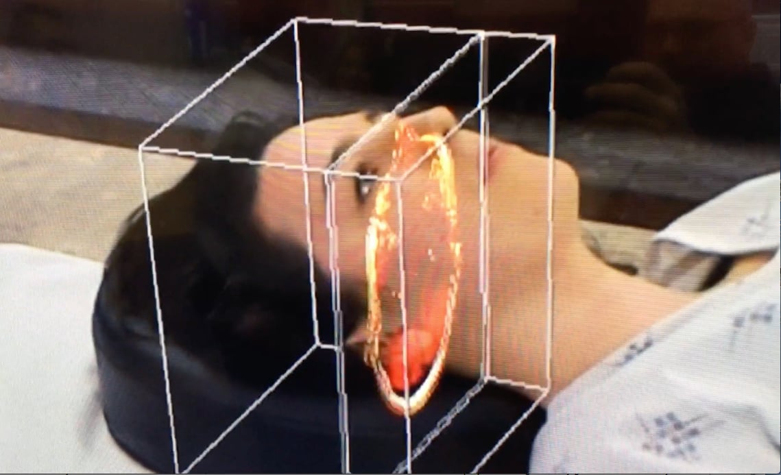 NovaRad showed its FDA-cleared OpenSight AR system for surgical planning. It integrates magnetic resonance imaging (MRI) or computed tomography (CT) imaging datasets into a HoloLens headset, and landmarks are registered to the patient on an operating table. The user can make hand gestures in midair to scroll through the image slices. The goal of the system is to allow for surgical planning.
Watch a VIDEO example of the NovaRad technology.
Biodigital showed its clinician educational AR system that allows users to view various organs in the body and manipulate or slice through the images via voice command and/or hand movements.
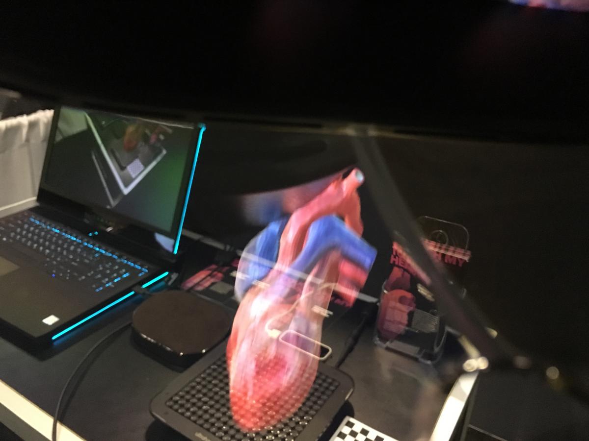 A really innovative example of AR is being developed by the company SoftServe. Its “Touch My Heart” work-in-progress technology allows anyone wearing an AR headset to not only see and interact with the heart, but also touch it. When the user moves their hand into the projected hologram, they get the sensation they are touching something. A pad below the image is composed of dozens of ultrasound transducers that emit sound waves in the shape of the heart so users feel touch sensations when their hand enters the virtual tissue.
Medivis unveiled its SurgicalAR platform for surgical applications. It uses a Microsoft HoloLens so physicians can overlay images directly onto the patient to aid in real-time procedural decision making. The imaging integrates with hospital picture archiving and communication systems (PACS).
Philips Healthcare is partnering with Microsoft on a work-in-progress mixed reality system designed for the interventional cath lab. Based on Philips’ Azurion angiography platform and Microsoft’s HoloLens 2 holographic computing platform, the new AR applications are designed for image-guided minimally invasive therapies. The companies showcased the technology for the first time at Mobile World Congress (MWC) in February.
At TCT, Abbott had an area in its booth where interventional cardiologists trained with VR visors in a virtual cath lab to use intravascular imaging catheters. Boston Scientific also is using this technology to train cardiologists.
Both GE Healthcare and TomTec at ASE showed work-in-progress VR systems to fire 3-D ultrasound imaging in true 3-D in order to better visualize the anatomy and to facilitate faster image manipulation workflow using hand movements. See these technologies in use in this VIDEO.
Watch the related VIDEO: Using Virtual and Augmented Reality to Examine Brain Anatomy and Pathology at MD Anderson.
Read the HIMSS 2019 article Virtual Reality Boosts Revenues and Patient Understanding.
2. Decrease on Hype and Focus on Introducing Artificial Intelligence Products
Two years ago at HIMSS 2017, you could not walk anywhere on the expo floor without vendor booth signage saying something about machine learning, artificial intelligence (AI), deep learning or "smart" software. This year 90 percent of those signs are gone as the hype cycle for AI comes to an end and attendees and hospitals expect to see real, actual products to purchase, not to have a theoretical discussion about how AI might help healthcare in the future. AI was still a big topic at HIMSS this year, but the focus is now on what is pending regulatory approval or FDA-cleared products.
AI is not a single product category — it comes in a multitude of flavors, performing scores of jobs across the healthcare spectrum and all subspecialties that were previously manual processes. While some offer first pass, automated diagnosis for STAT imaging like stroke, or to look for incidental findings flagged for the radiologist, most AI apps touted at HIMSS are in the backend of IT systems to speed workflow.
Two great examples of how AI is being leveraged to improve and speed up electronic patient record review were shown by Philips and Siemens. Both use AI to search for patient data and imaging relevant to specific care areas and simplify the view of the data into a patient timeline and dashboard so the clinicians do not have to sort through a sea of unrelated information and exams.
Philips showed its new Intellispace Oncology software, which uses a timeline and dashboard for a quick overview of a cancer patient on a single screen. The top of the screen has a timeline showing all patient encounters related to their cancer, including imaging exams, labs, pathology, procedures, etc. All are clickable and show a small pop-up box with a key image and brief description, and a link to the full set of images and the full report. Other boxes on the screen help organize the relevant patient data into easily digestible bits with links to full reports or areas of the patient record. An analytics button at the bottom of the page opens a window showing charts for things like tumor follow-up assessments to quickly show if the patient is responding to treatment.
Watch a quick VIDEO example of the Philips' software.
Siemens demonstrated its AI-Pathway Companion, which is currently under development. It uses a similar patient timeline and dashboard of relevant data and imaging, but has the added dimension of clinical decision support. In the case of a cancer patient, the AI will look at where the patient is at in terms of care and suggest next steps along the clinical pathway, including tests or imaging exams to order, or treatment options. The decision support is entirely based on medical society-set guidelines for evidence-based care. The goal of the technology is to help clinicians adhere to guideline-based care and offer information to referring physicians who might not know the standard of care with some forms of cancer, and the system will save them time otherwise spent researching information.
Watch a quick VIDEO example of the Siemens' technology.
Siemens also showed its AI-Rad Companion Chest CT, which is pending U.S. Food and Drug Administration (FDA) 510(k) clearance. It will be the first intelligent software assistant from Siemens for radiology. It is designed to identify anatomies on computed tomography imaging and potentially identify disease-relevant changes, differentiate between various structures and highlight them individually, and mark and measure potential abnormalities. This applies to the lungs, heart, aorta and coronary arteries. The software is designed to automatically turn findings into a quantitative report.
Fujifilm's REiLI artificial intelligence application for radiology was demonstrated, showing how it can auto-identify and -quantify abnormalities on medical images. One example shown was of a pleural effusion and fibrosis that the system color-coded, highlighting the areas of detected anomalies.
This topic is also discussed in the HIMSS19 VIDEO: What to Look for in Enterprise Imaging Systems.
3. Analytics is the Next Big Trend Following Digital Health Records Implementation
With the U.S. federal mandate under healthcare reform that all healthcare facilities convert from paper-based systems to electronic medical records (EMRs), it has effectively connected nearly all aspects of healthcare systems into a digital format over the past decade. This has made many hospitals focus efforts on leveraging this data by implementing powerful new analytics software. Many expected clinical decision support or other anticipated federal mandates to become the next big tree in health IT, but clearly providers have made the next/current major trend analytics to leverage their EMR investments.
Pulling data in the past was a tedious process and numbers were usually not available in real time. However, the recent generation of analytics software offered by all major health IT vendors has greatly simplified analytics and often allows for customizable dashboards to view all aspects of the care continuum. This includes the time it takes for patients to go through various procedures based on the number and specific types of procedures performed at each facility, equipment utilization or downtime, workload by clinician, patient length of stay based on ICD-10 codes, hospital-acquired infection based on department, and the list goes on and on.
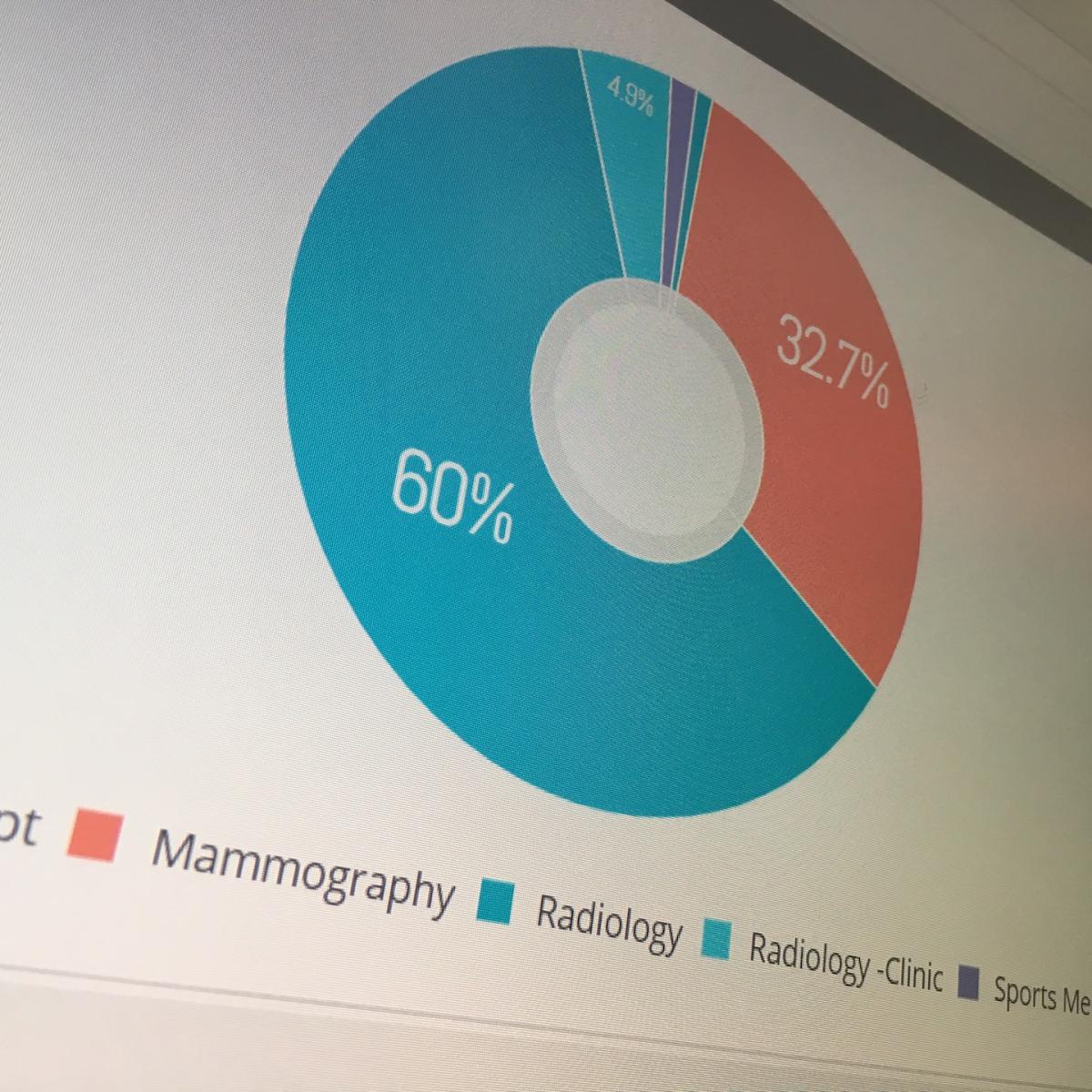 An example of this type of software at HIMSS 2019 was GE Healthcare's new analytics software for radiology business management. It offers a view over the entire department, but enables granular drilldown to analytics for specific machines, protocols, rooms and technologists to find bottlenecks and inefficiencies. The data can be presented in a numeric, line graph or pie graph format.
4. Creating Virtual Organs From Medical Imaging to Test Devices Before Implant
A really interesting work-in-progress technology shown by Siemens Healthineers was creating digital copies of patient's organs to preplan device implantations and assess if there is improved function or outcomes in a virtual environment before a patient enters a cath lab or operating room.
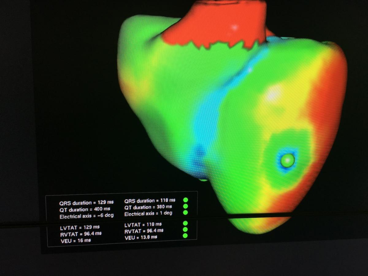 The company showed its first prototype "digital twins" software to create virtual hearts. It creates a digital organ that has the same electrophysiology characteristics as the patient’s real heart using a patient’s electrocardiogram (ECG), MRI scan and other data. Development is currently aimed at optimizing cardiac resynchronization therapy (CRT) lead placement. CRT currently has a 30 percent nonresponder rate, which is mainly due to the placement of leads. This model allows virtual placement of the leads In various locations to test response prior to the implantation procedure. Siemens said the technology also might have applications for virtual ablations.
See a VIDEO example of the virtual heart.
The vendor said the digital twin heart can exactly mirror the behavior of the patient’s real heart, displaying the same electrical activity, contraction, ejection fraction and pressure dynamics.
Siemens said smart algorithms could “learn” the function of virtually any organ in the body, potentially by mining data from patient images and their electronic medical record data. This could be a new direction for personalized healthcare that leverages existing patient imaging and data.
Similar work-in-progress technologies have been shown in the past year by other vendors for the virtual placement of stents in coronary arteries or virtual placement of heart valves to assess the hemodynamic function pre-procedure. These advances are image-based, complex virtual physiologic assessments that are being made being made possible by new computer algorithms, some of which include elements of artificial intelligence.
Read more about the virtual twin organ technology.
5. Cybersecurity Tops Concerns With Electronic Medical Records
As healthcare becomes more wired and interconnected, cybersecurity has become a primary concern of hospitals and it was a major topic at HIMSS 2019. Healthcare facilities have been the target of many high-profile attacks that have cost millions, open the facilities to liabilities and can cause a major disruption in patient care it systems are shut down or data is blocked by hackers.
While most people think security of digital systems is limited to patient data in the EMR, experts at HIMSS said some of the biggest threats to hospitals are the back-door computer systems that are not encrypted or updated for security. The first major EMR hacking incident a few years ago occurred after hackers accessed the hospital network through a wireless inventory connection with a soda vending machine. For this reason, many HIMSS vendors highlighted their security software for ancillary devices that are connected to the EMR, PACS or hospital information systems (HIS). These devices include ECG machines, imaging scanners, infusion pumps, telemetry systems, contrast injectors and anything else that interfaces with wired, wireless or Bluetooth connections.
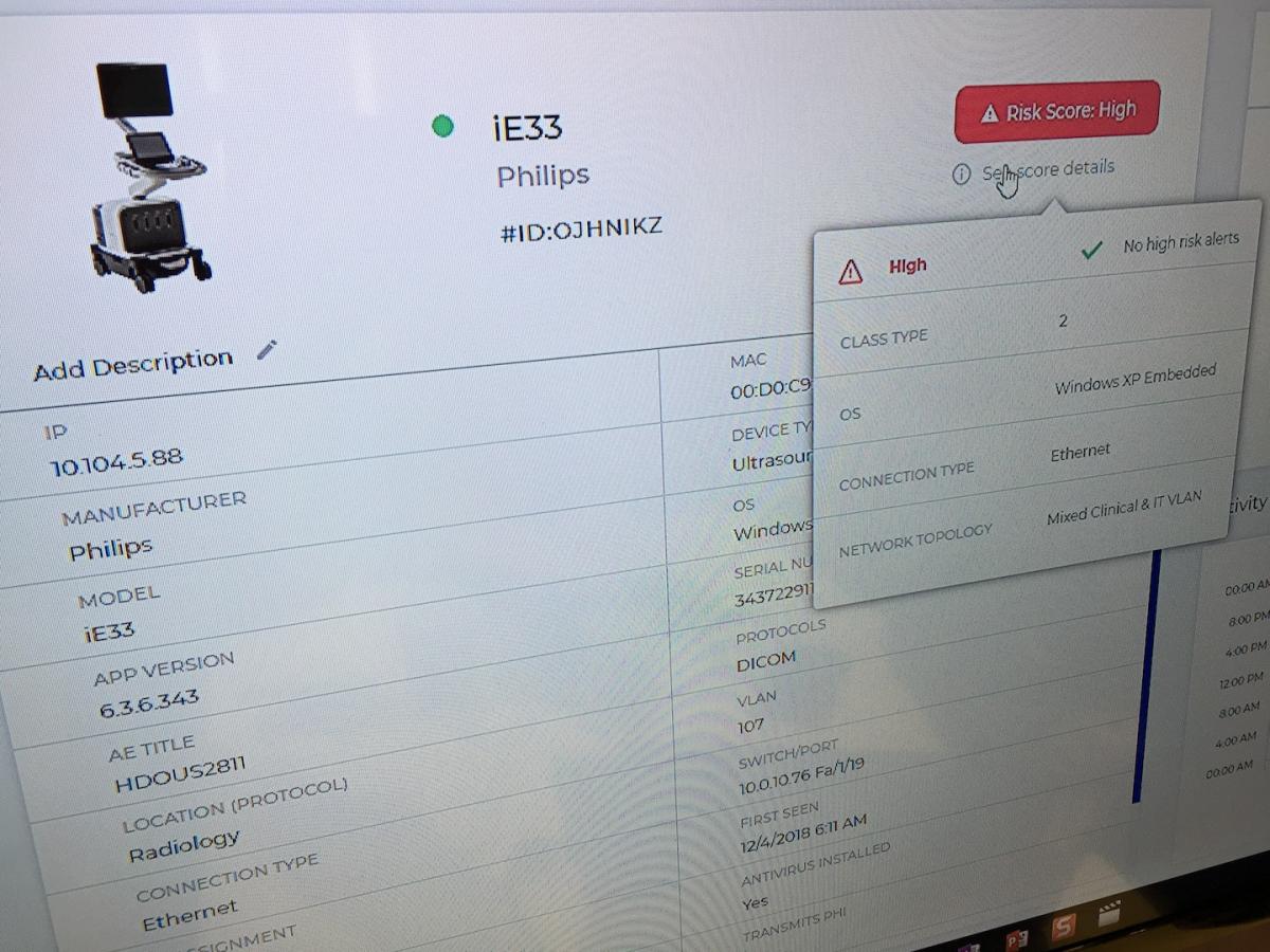 An example of this technology was shown by cybersecurity firm Medigate. When installed on medical device networks, the platform can detect early signs of attack and offer security risk assessments to help harden vulnerable equipment, or disconnect it from the network. The company has identified tens of thousands of devices for dozens of healthcare systems, many in the U.S. and Canada. Corporate partners include Palo Alto Networks and Cisco. The company said security flaws are often found in electronic medical device operating systems that have not been adequately patched, were not installed, or the systems are obsolete and no longer have any tech support
Read more about device cybersecurity at HIMSS.
6. Integrating Wearable Devices Into Patient Care
There is a good chance most patients already use some sort of wearable health device, including Apple Watches, Fitbits, Garmins and many others that have flooded the consumer market. However, while these devices offer data that might be of value to doctors caring for those patients, there have been few examples of how to integrate the technology into the current healthcare model.
"When tracking health data with wearables begins to integrate with healthcare organizations, it will be a tsunami of data," explained Karl Poterack, M.D., medical director, applied clinical informatics, Mayo Clinic, who spoke in sessions about wearables. "Fitbit alone has data on 3 trillion steps, and that is just a portion of the data they collect. There has to be a place to put this data and we need to answer the question of who owns it, and how it is collected and what happens to it when it goes to hospitals, which all have a different sent of rules on data collection under HIPAA."
He said a massive infrastructure would have to be built to address these concerns and handle the volume of wearable data that is out in the consumer world. However, no one will make the investment until research establishes guidelines or benchmarks for this data. This might include what it means if a cardiac patient has 1,000 steps as opposed to 15,000 steps a day.
"The larger barrier to wearables is what is the healthcare system going to do with that data," Poterack said. "The challenge is really how to take this data and link it to outcomes."
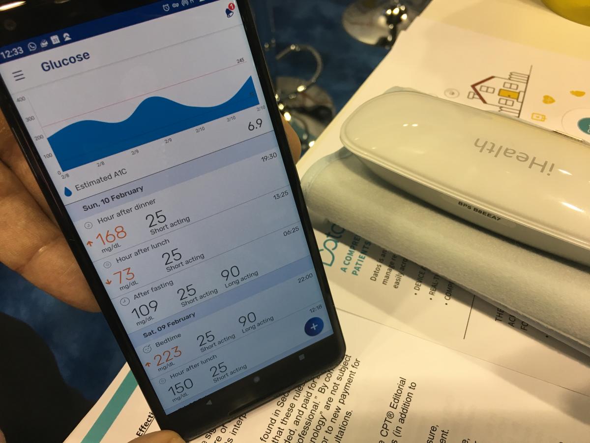 He and other speakers at HIMSS said major questions remain on how to interface wearable data into a usable format that is easily digestible for clinicians and the electronic medical record. An excellent example of how wearables, health apps and Bluetooth medical monitoring devices might be integrated into mainstream patient care is being piloted by Sheba Medical Center in Israel, one of the largest hospitals in the Middle East. Sheba had a booth on the HIMSS floor to highlight its health IT innovations, including its use of wearables to remotely monitor hundreds of patients each week in its cardiac rehabilitation program using consumer-grade devices.
Robert Klempfner, M.D., director of the Sheba Cardiovascular Prevention Institute, said patient compliance rates increased with the use of wearables, and the remotely monitored patients are no longer required to drive or take public transportation to the hospital for in-person sessions. He said the hospital also remotely monitors patients in its heart failure program using wearables as well.
Watch a VIDEO interview with Klempfner about the program at HIMSS 2019.
The hospital was able to interface an array of wearable devices and Bluetooth-enabled blood pressure cuffs, scales and glucose monitors into a single mobile phone app from the vendor Datos. The company can integrate data from a wide variety of wearable devices from several makers into a mobile app that can transfer the information to an electronic medical record. The app also offers two-way communication between the patient and the doctor’s office. The app has analytics on a patient’s health data, including charts and graphs showing patient progress.
Watch a short VIDEO example of this remote monitoring app technology.
Look through a photo gallery of other new technologies at HIMSS19.
Read the HIMSS19 article "Innovation to Address Physician Burnout."


 August 29, 2024


