November 8, 2017 — Konica Minolta Healthcare Americas Inc. presented its ultrasound systems and wireless digital radiology (DR) imaging technologies, both optimized for rheumatology practices, at the ACR/ARHP Annual Meeting, Nov. 3-8 in San Diego. The company sponsored several workshops dedicated to ultrasound education for rheumatologists, and also announced the availability of high definition (HD) and high dynamic range (HDR) X-ray imaging with AeroDR HD.
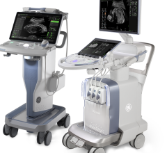
The Rheumatology Reporting Package for the SonImage HS1 delivers an efficient way to track the disease activity score of a patient and organize ultrasound studies, including storing still images with real-time clips. This integrated, customizable protocol can be operated with a foot switch, allowing the operator to move quickly through the exam without taking their hands off the probe.
The HS1 System also features Simple Needle Visualization (SNV) software to enhance needle clarity. The advanced algorithm is based on the movement of the needle and surrounding tissue, improving visibility for both in-plane and out-of-plane needle approaches. The resulting clarity of the needle, especially in steep angle approaches, enables increased accuracy in needle placement, making the portable ultrasound system an ideal solution for pain management guided injections, according to Konica Minolta.
The SonImage HS1 Compact Ultrasound System also offers advanced musculoskeletal (MSK) functionality to deliver high image quality and contrast resolution for diagnosis and management of rheumatic diseases. Ultrasound plays an important role in detecting abnormalities and assessing inflammatory joints, making the imaging modality more sensitive than clinical examination alone.
The HL18-4 wide-band linear probe available with SonImage HS1 provides real-time assessment of joint and tendon movements to aid in the detection of structural abnormalities. The hockey stick probe easily reaches difficult-to-access areas with its small footprint and maneuverability. The clinician can evaluate joints in the fingers and ankle more readily with the probe’s angulation, improved control and greater contact with the anatomy. The high-frequency probe provides high resolution in the near field for tissue differentiation and visualizes color flow with high Doppler sensitivity.
Konica Minolta sponsored 11 workshops at the ACR/AHRP Annual Meeting dedicated to providing training and education in the area of musculoskeletal ultrasound.
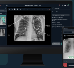
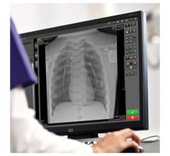
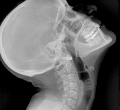
In addition to advanced ultrasound solutions, Konica Minolta also offers high definition systems for rheumatology practices, including the AeroDR HD X-ray flat panel detectors (FPD) that offer the option to switch between high definition and high dynamic range imaging. High definition imaging can be particularly useful over standard imaging when investigating small bone and joint structures. Using the high definition capability of AeroDR HD, it is easier to see the fine details to confidently diagnose the smallest anatomies. Switching to high dynamic range imaging on the AeroDR HD panel aggregates the data for a wider range of grays, providing a smooth image that allows clinicians to detect subtle differences in soft tissue.
AeroDR solutions now include Realism, Konica Minolta’s image processing that provides superior anatomy visualization within soft tissue and bony structures, according to the company. Realism’s enhanced sharpening and contrast sensitivity produces excellent detail of fine structures while optimizing image quality in low density areas, making images easier to read and improving productivity.
For more information: www.konicaminolta.com/medicalusa


 March 19, 2025
