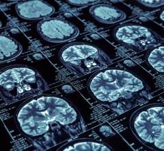
Brain imaging scans may one day provide useful information on the response to psychotherapy in patients with depression or anxiety, according to a review of current research in the November/December issue of the Harvard Review of Psychiatry, published by Wolters Kluwer.
Studies show promising initial evidence that specific "neuroimaging markers" might help in predicting the chances of a good response to psychotherapy, or choosing between psychotherapy or medications, in patients with major depressive disorder (MDD) and other diagnoses. "While some brain areas have emerged as potential candidate markers, there are still many barriers that preclude their clinical use," comments lead author Trisha Chakrabarty, M.D., of University of British Columbia, Vancouver.
Evidence of Neuroimaging Markers of Response to Psychotherapy
The researchers analyzed previous research evaluating brain imaging scans to predict the outcomes of psychotherapy for major depressive and anxiety disorders. Psychiatrists are interested in identifying brain imaging markers of response to psychotherapy — comparable to electrocardiograms and laboratory tests used to decide on treatments for myocardial infarction.
The review found 40 studies including patients with MDD, obsessive-compulsive disorder, posttraumatic stress disorder, and other diagnoses. Some studies used structural brain imaging studies, which show brain anatomy; others used functional scans, which demonstrate brain activity.
Although no single brain area was consistently associated with response to psychotherapy, the results did identify some "candidate markers." Studies suggested that psychotherapy responses might be related to activity in two deep brain areas: the amygdala, involved in mood responses and emotional memories; and the anterior insula, involved in awareness of the body's physiologic state, anxiety responses, and feelings of disgust.
In MDD studies, patients with higher activity in the amygdala were more likely to respond to psychotherapy. In contrast, in some studies of anxiety disorders, lower activity in the amygdala was associated with better psychotherapy outcomes. Studies of anterior insula activity showed the converse: psychotherapy response was associated with higher pretreatment activity in anxiety disorders and lower activity in MDD.
Other studies linked psychotherapy responses to a frontal brain area called the anterior cingulate cortex, which plays a critical role in regulating emotions. Most of the evidence suggested that MDD patients with lower activity in some parts of the ACC (ventral and subgenual) were more likely to have good outcomes with psychotherapy.
"Future studies of psychotherapy response may focus further on these individual regions as predictive markers," according to Chakrabarty. "Additionally, future biomarker studies may focus on pretreatment functional connectivity between these regions, as affective experience is modulated via reciprocal connections between brain areas such as the ACC and amygdala."
The researchers emphasize the limitations of current evidence on neuroimaging markers of psychotherapy response — the studies were highly variable in terms of their methodology and results. Further studies are needed to assess how the potential neuroimaging markers perform over time, whether they can predict which patients will respond better to medications versus psychotherapy, and how they might be integrated with clinical features in order to improve outcomes for patients with depression and anxiety disorders.


 July 25, 2024


