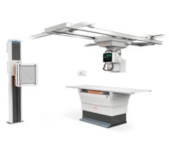
August 8, 2016 — Konica Minolta announced the release of its newest X-ray digital radiography (DR) system at the 2016 Association for Medical Imaging Management (AHRA) annual meeting, July 31-Aug. 3 in Nashville. Combining a built-from-the-ground-up design along with the fast and advanced Ultra image processing software, the company said the system (name not yet revealed) offers unprecedented workflow, clinical, patient convenience and satisfaction benefits.
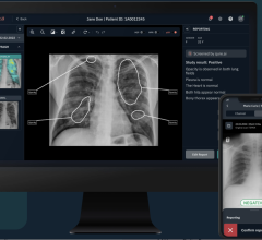
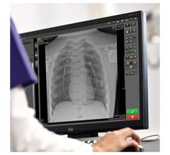
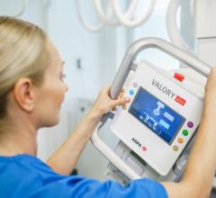
The DR U-Arm system is a floor-mounted system that provides numerous benefits including faster exams, smoother positioning and improved stitching processes for advanced studies. The Ultra software uses a full console screen right at the point of patient contact for accepting or rejecting images so the technologist can remain with the patient without returning to the workstation. With the ability to view results at the tube stand, X-ray exams are quicker and more comfortable for patients, and enable increased throughput for the facility.
In addition to the speed and efficiency benefits for the technologist and patient, the new weight-bearing stand that sits only 10 inches from the floor provides benefits as well. The low step-up height creates an easy-to-use imaging environment for patients challenged by mobility, be it athletes, diabetics or obese patients. The low-to-the-ground design also brings benefits for those in wheelchairs — who can remain seated in their wheelchair for image acquisition — and for pediatric populations who are often calmer and more comfortable when they are lower.
Contributing further to patient comfort is the hardware design of the U-Arm, which enables AP and oblique imaging and then lateral positioning with one push of a button, allowing the patient to stay on the stand for the whole exam.
The U-Arm also creates a level of efficiency that changes the way imaging patients flow through the health system, according to Konica Minolta. While hospitals are struggling to keep medical imaging exams within their walls, the company said it offers a technology that is versatile enough to serve patients not only in radiology but from urgent care to specialty areas like orthopedics, pediatrics and rheumatology. And with an aesthetic design that is compact, the system will fit in older facilities challenged with low, 8-foot ceilings.
Konica Minolta rounded out its exhibit at AHRA by also highlighting its Exa software platform for enhanced image sharing, viewing and storage, along with its hand-carried ultrasound system, the SonImage HS1.
- Exa PACS – A Web-based platform that the company says was the first picture archiving and communication system (PACS) to enable zero footprint full-featured diagnostic viewing, Exa works on any consumer-grade PC with any operating system without plug-ins or installations. It boasts a configurable dashboard and custom workflow design, as well as advanced dictation and viewing tools.
- Exa Mammo Viewer – Available either as a standalone mammography viewer or as part of the Exa PACS platform, Exa Mammo leverages the benefits of the existing Exa platform to enable anywhere viewing for all breast imaging modalities including 2-D or 3-D mammography studies and digital breast tomosynthesis. With the benefit of server-side rendering, radiologists have fast access with a seamless DICOM-compliant PACS to breast images at any time from any location.
- SonImage HS1 – A hand-carried ultrasound system featuring a compact design that is particularly well-suited for musculoskeletal exams.
For more information: www.konicaminolta.com/medicalusa

 July 18, 2024
