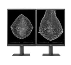
As breast density increases, mammography sensitivity decreases and breast cancer risk elevates, highlighting the need for optimal approaches to individualized breast cancer screening.
As radiologists, most of us have seen firsthand how dense breast tissue can mask cancer in mammography. As breast density increases, mammography sensitivity decreases and breast cancer risk elevates, highlighting the need for optimal approaches to individualized breast cancer screening. In women with dense breast tissue, this is particularly challenging as it can equate to the perfect storm, where mammography is less effective and the risk of breast cancer is increased.
In May, Vermont became the 28th state to pass legislation requiring that women with dense breasts receive dense breast notifications (DBNs) following mammography. As more states continue to pass this legislation, DBNs are rapidly becoming a standard component of breast cancer screening and overall patient care.
While this is a very positive step in women’s health, a recent study published in the Journal of the American Medical Association (JAMA) suggests that some patients may not understand the language used in DBNs received following mammography.1 The study authors also noted that the content of these letters can vary from state to state, the language used exceeds the average American’s reading level, and these communications do not always provide clear next-steps for patients who might benefit from additional screening. Some require that women be informed if additional screening can detect mammographically occult breast cancer, and some only require that women be informed of what their breast density is, without context to what the implications and possible strategies for dense breast tissue are.
This above mentioned study identified a critical area where the medical community can, and should, improve patient care. While critics of this legislation have stated that these laws can cause confusion, anxiety and added costs, advocates of these laws feel DBNs deliver crucial information that can empower patients to make more informed decisions about their own healthcare. Empowering women to insist on optimizing their health and particularly their breast health is critical, and in fact, is one of the fundamental principles of the Brem Foundation to Defeat Breast Cancer, a cause near and dear to me.
A Call to Action
We must take steps to enhance our communications with patients related to breast density to ensure they understand their risk for developing breast cancer and whether additional screening may be warranted. We cannot wait for national legislation to introduce uniform standards for DBNs.
It should be noted that these communications are intended to initiate a dialogue between patient and doctor, rather than replace it. It is important for doctors to follow up appropriately after mammography to ensure that patients are well informed about their breast density, their potential risk for developing cancer and whether they might benefit from additional screening.
Radiologists involved in interpretation of mammography are the ones charged with determining breast density. It is our responsibility to communicate these results to patients and their care teams. Though it is not required by law, at George Washington University Hospital we inform patients and their referring physicians about their breast density following mammography.
Advanced Solutions
Regardless of how we communicate with our patients about breast density, the bottom line remains that DBNs can only be as effective as the accuracy of the breast density assessment itself. The American College of Radiology recommends the BI-RADS scale as a standardized system to categorize breast density. However, this standardized system is subjective and both breast imagers and general radiologists report that reader variability is a challenge when determining breast density.2 This is because most radiologists determine breast density in the subjective, BI-RADS manner. However, an objective, true 3-D approach to determining breast density could standardize the determination of breast density and thereby make the process more straightforward for radiologists and patients.
The advent of advanced software programs will help to address the issue of reader variability by providing more consistent breast density assessments that are accurate and reproducible. This technology automates the same analytical approach used by experienced radiologists, as it analyzes digital mammograms, calculates the patient’s breast density and determines the appropriate density category corresponding to BI-RADS standards. The increased use of new software also can help radiologists with workflow while more precisely identifying patients who would benefit from additional screening.
Another recent study adds to the growing body of evidence that supports the use of tomosynthesis in breast cancer screening, both for women with dense breasts and non-dense breasts.3 Tomosynthesis is rapidly being adopted in clinical practice because of its advantages, including decreased callback rates and slight increase in cancer detection. But radiologists often report that the interpretation of tomosynthesis requires more time, as compared to 2-D mammography. Much-needed computer aided detection (CAD) technology is being developed to help radiologists manage the increased workflow and reduce the time required for interpretation of tomosynthesis by assisting the radiologist with the identification of potential mammographic abnormalities, without sacrificing reader performance. An example of this type of software is iCAD’s concurrent read tomosynthesis CAD tool, which analyzes hundreds of breast images, identifies potential areas of interest and puts the images together into a synthetic 2-D image for the radiologist. The enhanced synthetic 2-D image is also linked to the 3-D tomosynthesis dataset that helps radiologists more quickly and efficiently read mammogram results.
The implementation of tomosynthesis, CAD solutions and other workflow tools would markedly increase the usability of tomosynthesis in clinical practice and allow for more accurate cancer detection in patients with both dense and non-dense breasts.
The Debate Continues
Another key issue is that the medical community has not agreed on a protocol regarding supplemental screening for women with dense breasts. Studies have demonstrated that ultrasound, magnetic resonance imaging (MRI) and breast specific gamma imaging (BSGI) can detect mammographically occult cancer. However, additional screening also finds benign lesions and results in increased biopsy rates. What is clear is that mammography is no longer a one-size-fits all solution. With the increase in the technologies available and the individualization of screening, the technologies used need to be determined by a woman’s risk. The approach of risk-based screening is a necessary one, with dense breast tissue being a stratification factor in screening approaches.
The increased adoption of advanced technology, along with improved DBNs and improved strategies for efficient and effective determination of breast density, in addition to implementation of risk-based screening with adjunct screening protocols in women with dense breast tissue as well as an approach to consistent, comprehendible dialogue with patients, can help the radiological community improve patient care. This can ultimately mean the difference between a diagnosis of early, curable breast cancer and detection of breast cancer in more advanced stages, when it is less treatable and potentially fatal.
References:
1 Kressin NR, Gunn CM, Battaglia TA. “Content, Readability, and Understandability of Dense Breast Notifications by State.” JAMA. 2016;315(16):1786-1788. doi:10.1001/jama.2016.1712.
2 Buist DSM, Anderson ML, Haneuse SJPA, et al. “Influence of Annual Interpretive Volume on Screening Mammography Performance in the United States.” Radiology. 2011;259(1):72-84. doi:10.1148/radiol.10101698.
3 Elizabeth A. Rafferty, Melissa A. Durand, Emily F. Conant, Debra Somers Copit, Sarah M. Friedewald, Donna M. Plecha, Dave P. Miller. “Breast Cancer Screening Using Tomosynthesis and Digital Mammography in Dense and Nondense Breasts.” JAMA, 2016; 315 (16): 1784.
Rachel Brem, M.D., is an expert in the field of breast imaging and intervention. She is the director of breast imaging and intervention, professor of radiology at The George Washington University Medical Center and program leader for breast cancer at the George Washington Cancer Center. Brem is also a founder and chief medical advisor for the Brem Foundation to Defeat Breast Cancer.


 March 21, 2025


