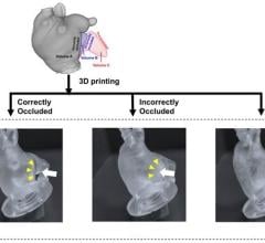
June 13, 2016 – While 3-D printing has been around since the mid-1980s, there is accumulating evidence that this technology has the potential to revolutionize the understanding and management of heart conditions. A team of researchers from the Cleveland Clinic created a 3-D model of an aortic valve from a patient with a severe case of calcific aortic stenosis (AS) — narrowing of the aortic valve — to simulate the patient’s beating heart and assess the blood flow, or “hemodynamics.”
“In order to better understand the physiology of AS (which can be complex), we produced a true replicate of the valve using 3-D printing technology. Then, to assess the valve hemodynamics, we placed the 3-D-printed valve into a circuit where flow can be controlled and we can try out different flow conditions. In this research, we present a proof of concept case of severe AS where the pressure gradients, obtained by cardiac ultrasound, were successfully replicated in the circuit built around the 3-D-printed version of the stenotic valve,” said lead author Serge Harb, M.D., of Cleveland Clinic, Cleveland, Ohio.
The hemodynamic results using this 3-D-printed valve in the flow circuit simulating the pumping action of the heart, were confirmed by Doppler echocardiography, the technique used in daily practice to evaluate patients with AS. Harb said, “This technology shows a lot of promise. Not only will it help us better understand the mechanisms of the disease, but it also has the potential to provide a more personalized treatment where the particular valve of the affected patient is 3-D-printed, guiding its optimal management. This may be particularly helpful for surgical planning, or when using new catheter-based technologies for non-surgical valve replacement.”
Researchers on the study, Three Dimensional (3D) Printing and Functional Assessment of Aortic Stenosis Using a Flow Circuit: Feasibility and Reproducibility, include Harb, Ryan Klatte, Brian P. Griffin and Leonardo L. Rodriguez, from the Cleveland Clinic Foundation, Cleveland, Ohio.
Harb presented a poster based on this research during the American Society of Echocardiography (ASE) 27th Annual Scientific Sessions, June 10-14 in Seattle.
For more information: www.asescientificsessions.org


 April 18, 2025


