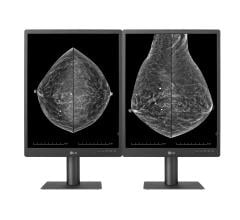
May 2, 2016 — In a new report, the U.S. Food and Drug Administration (FDA) suggests that since the passage of the Mammography Quality Standards Act (MQSA), proper patient positioning remains one of the most important aspects of high-quality mammography imaging.
Passed in 1999, the MQSA is the primary mechanism for enforcing compliance with quality standards for mammography facilities. The FDA reports that as of May 1, 2016, there were a total of 8,738 certified facilities in the United States sporting a total of 16,042 accredited units. Of those certified facilities, 8,493 had full field digital mammography (FFDM) units; the FDA reported a total of 15,744 accredited FFDM units [1].
The agency notes that positioning is important because only those portions of the breast which are included on the mammographic image can be evaluated for signs of cancer. Any portion of the breast which is not imaged cannot be evaluated, and cancers in those portions of the breast can be missed. In a 2002 study, the "[s]ensitivity [of mammography] dropped from 84.4 percent among cases with passing positioning to 66.3 percent among cases with failed positioning" [2].
Poor positioning has been found to be the cause of most clinical image deficiencies and most failures of accreditation, according to the FDA. In 2015, the American College of Radiology (ACR), the largest FDA-approved accreditation body (AB), found that of all clinical images which were deficient on the first attempt at accreditation, 92 percent were deficient in positioning. Also, in ACR-accredited facilities, 79 percent of all unit accreditation failures in 2015 were due to positioning.
The FDA reported that similar results were noted by the Iowa and Texas state ABs: in 2015, positioning was a cause of 91 percent of clinical image failures in Iowa and 100 percent of clinical image failures in Texas.
MQSA requires that the "[c]linical images produced by any certified facility must continue to comply with the standards for clinical image quality established by that facility's accreditation body" [3]. Positioning failures of clinical images often lead to an Additional Mammography Review (AMR). If the AMR also reveals failing results, the FDA will determine whether the facility’s practice of mammography represents a serious risk to health, and may order a facility to cease performing mammography and to notify all affected patients and their referring healthcare providers about the facility’s image quality problems. Thus, the consequences of poor positioning can be very significant not just for individual patients, but for mammography facilities as well.
Although the technologist performs the mammogram, the responsibility for correct positioning is shared by the technologist and the interpreting physician. This shared responsibility is reflected in MQSA. The Preamble to the MQSA Final Rule emphasizes that all interpreting physicians are "the final arbiters of the quality of mammography images," and adds, "It is important that they communicate their satisfaction or dissatisfaction with the quality of the images they are provided to interpret to the technologists who produced them. Such communication is the crucial first step in the identification of problems and the initiation of corrective actions" [4].
Thus, to achieve and maintain proper positioning, both training and communication are essential, according to the FDA. Technologists should be trained in proper positioning, and should seek feedback from fellow technologists and interpreting physicians. Interpreting physicians, in turn, should review the elements of proper patient positioning, and give constructive feedback to technologists on the positioning of the mammograms that are presented to them for interpretation.
References
- “MQSA National Statistics,” MQSA Insights, www.fda.gov.
- Taplin SH, Rutter CM, Finder C, et al. Screening Mammography: Clinical Image Quality and the Risk of Interval Breast Cancer. AJR 2002; 178: 797-803.
- 21 CFR § 900.12(i).
- 62 Fed. Reg. 55935 (Oct. 28, 1997).


 July 29, 2024


