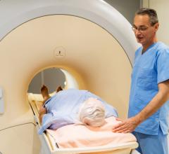
April 22, 2015 — Although ultrasound remains the primary imaging modality used in prenatal imaging, fetal magnetic resonance imaging (MRI) is playing an increasing role in further evaluation of fetuses suspected of congenital anomalies. As 3-T MRI scanners become more common due to their improved image signal-to-noise ratio and anatomical detail, the benefits of 3-T MRI must be weighed against potential risks to the fetus that may result from the higher field strength.
"MRI is playing an increasingly important role in the assessment of complex prenatal disease," said Kathleen E. Carey, M.D., Mayo Clinic, Jacksonville, Florida. "The use of stronger 3-T field strengths may allow for improved visualization of subcutaneous fat and osseous structures, including the hands and feet of the developing fetus."
The study is featured in an electronic exhibit at the American Roentgen Ray Society (ARRS) 2015 annual meeting in Toronto.
First attempted in the 1980s, MRI in pregnancy has maintained a steady growth rate. Recent studies have demonstrated that fetal MRI not only provides good detail of normal fetal anatomy and allows characterization of suspected anomalies but also may alter the diagnosis provided by ultrasound, uncovering fetal anomalies earlier and more accurately. Currently, clinical MRI is most frequently performed on 1.5-T MRI systems, but 3-T scanners are becoming more common around the world as the search for improved image signal-to-noise ratio and anatomic detail grows. This developmental opportunity must be weighed against potential risks to the fetus, taking into account the specific absorption rate.
ARRS will present a series of fetal MRI cases and discuss the anatomic and imaging findings, indication for MRI, diagnosis and impact on patient management. The benefits and limitations of fetal MRI at 1.5-T and 3-T field strengths will be discussed, and future advanced imaging will be considered, including spectroscopy and diffusion tensor imaging.
For more information: www.arrs.org

 April 10, 2025