Angiographic imaging system vendors have developed several new technologies to address emerging cath lab trends, including the need to reduce radiation dose, improve image quality and enable advanced procedural image guidance. All three of these points have become increasingly important as more complex procedures are attempted in interventional labs and hybrid ORs. These procedures include embolic coiling, neuro-interventions, thrombectomy, aortic repair, transcatheter aortic valve replacement (TAVR), mitral clip valve repairs, left atrial appendage (LAA) occlusions, atrial and ventricular septal defect closures, and new interventions for both electrophysiology (EP) and heart failure.
All of the major vendors in the United States recently introduced new systems and technologies to reduce dose and enhance visualization in the interventional lab. The vendors have tailored their systems into various models for specific specialties and at various price points depending on the degree of functionality.
Advanced Imaging, Guidance, Planning
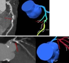
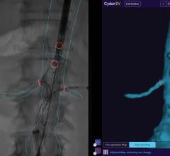
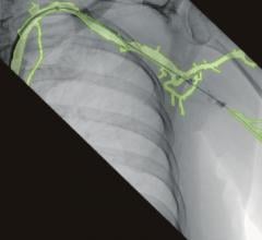
Newer imaging systems enable advanced 3-D imaging with rotational angiography, which uses a quick spin around the patient to create a computed tomography (CT)-like, 3-D image of the anatomy. This can all be done tableside in the lab. Some systems allow these images, or CT or MRI (magnetic resonance imaging) 3-D images, to be overlaid or fused with the live 2-D fluoroscopic images. This fusion technology is used with TAVR planning and navigation software to better guide precise device placement. Software also allows 3-D images to be integrated with EP electromapping systems to guide catheter ablation procedures without the need for live fluoroscopy, helping to reduce dose.
Radiation Reduction
Ionizing radiation from medical imaging has become a growing concern, but new technologies can help reduce dose. In the lab, as transcatheter procedures become more complex and require longer imaging times, radiation exposure of both patients and operators is increasing.
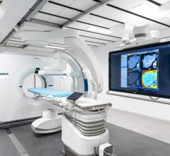
Newer, large-format displays now available for cath labs enable a larger field of view and better visualization of the anatomy, instead of operators leaning over the table to try to see anatomic details on smaller screens, said John Messenger, M.D., FACC, director, cath labs, University of Colorado, who spoke on new angiography technologies at the 2014 American College of Cardiology (ACC) meeting. He said this helps better visualize procedures and lower dose by not needing to image the anatomy several times.
Messenger said all the major vendors recently released new angiography systems with dose-lowering technologies. These advances include new X-ray tubes and more sensitive detectors and software to help improve image quality at lower doses, in addition to noise reduction. He pointed to the Philips Allura Clarity as an example of next-generation angiography system that can help lower standard procedural dose by 50 to 75 percent. He said the operator can easily adjust the frame rates to help reduce dose, and the system incorporates Philips’ ClarityIQ software to achieve excellent visibility at low X-ray dose levels for patients of all sizes. The software helps correct for motion, reduces noise, auto enhances the image and corrects pixel shift on cine images. The ClarityIQ technology is also available as an upgrade for the majority of Philips’ installed base of monoplane and biplane interventional X-ray systems.
He said another recent innovation to help lower dose is Toshiba’s Spot Fluoroscopy software for its Infinix-i systems. It allows clinicians to observe a target region of anatomy using Spot Fluoro’s live fluoroscopy, while viewing the last image hold (LIH) in the surrounding area that has been collimated out of the field of view to cut dose.
Newly Released Systems
Siemens introduced several new systems in its Artis series, including the Artis Q, Artis Q.zen and the Artis one. These incorporate both new X-ray tube and detector technology to not only lower dose, but also cut energy consumption by up to 20 percent from previous generation Artis systems. Siemens’ new X-ray tube uses flat emitter technology, which enables smaller, square focal spots that the vendor said leads to as much as 70 percent improved visibility of small vessels and improved image quality. The Artis Q.zen combines this X-ray tube with a new crystalline detector that enables patient doses as low as half the standard angiography levels.
The floor-mounted Artis one gained U.S. Food and Drug Administration (FDA) clearance in May. It features positioning flexibility similar to ceiling-mounted systems and occupies an area of 269 square feet, compared to the 484-square-feet required by ceiling-mounted systems. It has several axes that can move independently, enabling easy positioning. It accommodates full head-to-toe coverage of patients up to 6 feet, 10 inches without the need for repositioning.
Shimadzu gained FDA clearance in March 2014 for its new Trinias series. It uses high-speed Score Pro image processing technology, and a smart ergonomic design aids intuitive functionality. The Score process enhances fluoroscopic images while maintaining as low as achievable dose considerations.
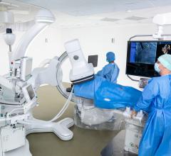
GE Healthcare introduced its Discovery IGS series in 2013. The laser-guided system captures the advantages of floor- and ceiling-mounted fixed systems and the mobility advantage of mobile C-arms without any of their inherent limitations. The imaging device is on a mobile, wheeled gantry that can be moved anywhere in the room like a mobile C-arm, but offers precision, high-quality imaging and features of a fixed system. At the touch of a button, clinicians can move the system to the table for imaging various anatomies and then move it aside and park it, allowing room to optimally position physicians, nurses, technologists, anesthesiologists and other personnel for surgery, with unobstructed access to the patient. The gantry comes with a new wide-bore design, which allows for steep angles and ease in 3-D acquisition, especially for large patients. It uses the same digital flat-panel detector technology as GE’s fixed-base Innova angiography imaging systems.


 May 17, 2024
